


Crystal Structure of D-alanine--D-alanine ligase from Klebsiella pneumoniae subsp. pneumoniae in complex with AMP
Abendroth, J., Bolejack, M.J., Lorimer, D.D., Horanyi, P.S., Edwards, T.E.To be published.
Experimental Data Snapshot
Starting Model: experimental
View more details
Entity ID: 1 | |||||
|---|---|---|---|---|---|
| Molecule | Chains | Sequence Length | Organism | Details | Image |
| D-alanine--D-alanine ligase | 384 | Klebsiella pneumoniae subsp. pneumoniae HS11286 | Mutation(s): 0 Gene Names: ddl, KPHS_10610 EC: 6.3.2.4 | 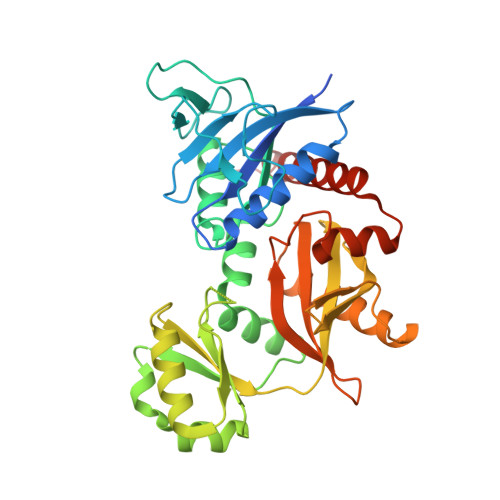 | |
UniProt | |||||
Find proteins for A0A0H3GSI5 (Klebsiella pneumoniae subsp. pneumoniae (strain HS11286)) Explore A0A0H3GSI5 Go to UniProtKB: A0A0H3GSI5 | |||||
Entity Groups | |||||
| Sequence Clusters | 30% Identity50% Identity70% Identity90% Identity95% Identity100% Identity | ||||
| UniProt Group | A0A0H3GSI5 | ||||
Sequence AnnotationsExpand | |||||
Reference Sequence | |||||
| Ligands 4 Unique | |||||
|---|---|---|---|---|---|
| ID | Chains | Name / Formula / InChI Key | 2D Diagram | 3D Interactions | |
| AMP (Subject of Investigation/LOI) Download:Ideal Coordinates CCD File | C [auth A], K [auth B] | ADENOSINE MONOPHOSPHATE C10 H14 N5 O7 P UDMBCSSLTHHNCD-KQYNXXCUSA-N |  | ||
| EDO Download:Ideal Coordinates CCD File | E [auth A] F [auth A] G [auth A] H [auth A] M [auth B] | 1,2-ETHANEDIOL C2 H6 O2 LYCAIKOWRPUZTN-UHFFFAOYSA-N |  | ||
| CL Download:Ideal Coordinates CCD File | I [auth A], J [auth A], Q [auth B], R [auth B] | CHLORIDE ION Cl VEXZGXHMUGYJMC-UHFFFAOYSA-M |  | ||
| MG (Subject of Investigation/LOI) Download:Ideal Coordinates CCD File | D [auth A], L [auth B] | MAGNESIUM ION Mg JLVVSXFLKOJNIY-UHFFFAOYSA-N |  | ||
| Length ( Å ) | Angle ( ˚ ) |
|---|---|
| a = 95.35 | α = 90 |
| b = 115.42 | β = 90 |
| c = 68.48 | γ = 90 |
| Software Name | Purpose |
|---|---|
| XDS | data reduction |
| XSCALE | data scaling |
| PHENIX | refinement |
| PDB_EXTRACT | data extraction |
| MoRDa | phasing |
| Coot | model building |
| Funding Organization | Location | Grant Number |
|---|---|---|
| National Institutes of Health/National Institute Of Allergy and Infectious Diseases (NIH/NIAID) | United States | HHSN272201700059C |


