7U4N

Crystal structure of human GPX4-U46C in complex with RSL3


Experimental Data Snapshot

  • Method: X-RAY DIFFRACTION
  • Resolution: 1.60 Å
  • R-Value Free: 0.188 
  • R-Value Work: 0.150 
  • R-Value Observed: 0.153 

wwPDB Validation   3D Report Full Report


Ligand Structure Quality Assessment 


This is version 1.2 of the entry. See complete history


Literature

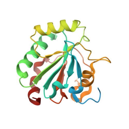
Small-molecule allosteric inhibitors of GPX4.

Liu, H.Forouhar, F.Lin, A.J.Wang, Q.Polychronidou, V.Soni, R.K.Xia, X.Stockwell, B.R.

(2022) Cell Chem Biol 29: 1680-1693.e9

  • DOI: https://doi.org/10.1016/j.chembiol.2022.11.003
  • Primary Citation of Related Structures:  
    7U4I, 7U4J, 7U4K, 7U4L, 7U4M, 7U4N

  • PubMed Abstract: 

    Encouraged by the dependence of drug-resistant, metastatic cancers on GPX4, we examined biophysical mechanisms of GPX4 inhibition, which revealed an unexpected allosteric site. We found that this site was involved in native regeneration of GPX4 under low glutathione conditions. Covalent binding of inhibitors to this allosteric site caused a conformational change, inhibition of activity, and subsequent cellular GPX4 protein degradation. To verify this site in an unbiased manner, we screened a library of compounds and identified and validated that an additional compound can covalently bind in this allosteric site, inhibiting and degrading GPX4. We determined co-crystal structures of six different inhibitors bound in this site. We have thus identified an allosteric mechanism for small molecules targeting aggressive cancers dependent on GPX4.


  • Organizational Affiliation

    Department of Chemistry, Columbia University, New York, NY 10027, USA.


Macromolecules
Find similar proteins by:  (by identity cutoff)  |  3D Structure
Entity ID: 1
MoleculeChains Sequence LengthOrganismDetailsImage
Phospholipid hydroperoxide glutathione peroxidase
A, B
192Homo sapiensMutation(s): 1 
Gene Names: GPX4
EC: 1.11.1.12
UniProt & NIH Common Fund Data Resources
Find proteins for P36969 (Homo sapiens)
Explore P36969 
Go to UniProtKB:  P36969
PHAROS:  P36969
GTEx:  ENSG00000167468 
Entity Groups  
Sequence Clusters30% Identity50% Identity70% Identity90% Identity95% Identity100% Identity
UniProt GroupP36969
Sequence Annotations
Expand
  • Reference Sequence
Small Molecules
Ligands 1 Unique
IDChains Name / Formula / InChI Key2D Diagram3D Interactions
L8X
Query on L8X

Download Ideal Coordinates CCD File 
C [auth A],
D [auth B]
methyl (1S,3R)-2-(chloroacetyl)-1-[4-(methoxycarbonyl)phenyl]-2,3,4,9-tetrahydro-1H-pyrido[3,4-b]indole-3-carboxylate
C23 H21 Cl N2 O5
TXJZRSRTYPUYRW-NQIIRXRSSA-N
Experimental Data & Validation

Experimental Data

  • Method: X-RAY DIFFRACTION
  • Resolution: 1.60 Å
  • R-Value Free: 0.188 
  • R-Value Work: 0.150 
  • R-Value Observed: 0.153 
  • Space Group: P 1 21 1
Unit Cell:
Length ( Å )Angle ( ˚ )
a = 32.899α = 90
b = 92.856β = 100.19
c = 53.786γ = 90
Software Package:
Software NamePurpose
PHENIXrefinement
PDB_EXTRACTdata extraction
XDSdata reduction
XDSdata scaling
MOLREPphasing

Structure Validation

View Full Validation Report


Ligand Structure Quality Assessment 


Entry History & Funding Information

Deposition Data


Funding OrganizationLocationGrant Number
National Institutes of Health/National Cancer Institute (NIH/NCI)United StatesP01CA87497
National Institutes of Health/National Institute of General Medical Sciences (NIH/NIGMS)United StatesR24GM141256
National Institutes of Health/National Institute of Neurological Disorders and Stroke (NIH/NINDS)United StatesR61NS109407
National Institutes of Health/National Cancer Institute (NIH/NCI)United StatesR35CA209896

Revision History  (Full details and data files)

  • Version 1.0: 2022-12-07
    Type: Initial release
  • Version 1.1: 2022-12-14
    Changes: Database references
  • Version 1.2: 2022-12-28
    Changes: Database references