7TX4

Neutron crystal structure of SARS-CoV-2 NSP3 macrodomain at 293 K (P21 crystal form)


Experimental Data Snapshot

  • Method: X-RAY DIFFRACTION
  • Resolution: 1.90 Å
  • R-Value Free: 0.224 
  • R-Value Work: 0.166 

  • Method: NEUTRON DIFFRACTION
  • Resolution: 2.35 Å
  • R-Value Free: 0.260 
  • R-Value Work: 0.177 

wwPDB Validation   3D Report Full Report


This is version 1.2 of the entry. See complete history


Literature

The mechanisms of catalysis and ligand binding for the SARS-CoV-2 NSP3 macrodomain from neutron and x-ray diffraction at room temperature.

Correy, G.J.Kneller, D.W.Phillips, G.Pant, S.Russi, S.Cohen, A.E.Meigs, G.Holton, J.M.Gahbauer, S.Thompson, M.C.Ashworth, A.Coates, L.Kovalevsky, A.Meilleur, F.Fraser, J.S.

(2022) Sci Adv 8: eabo5083-eabo5083

  • DOI: https://doi.org/10.1126/sciadv.abo5083
  • Primary Citation of Related Structures:  
    7TWF, 7TWG, 7TWH, 7TWI, 7TWJ, 7TWN, 7TWO, 7TWP, 7TWQ, 7TWR, 7TWS, 7TX3, 7TX4, 7TX5

  • PubMed Abstract: 

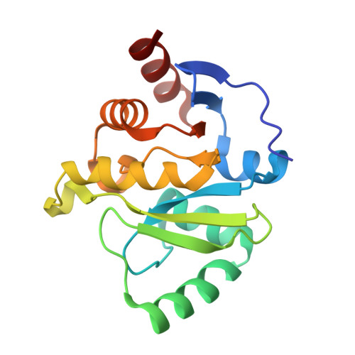
    The nonstructural protein 3 (NSP3) macrodomain of severe acute respiratory syndrome coronavirus 2 (SARS-CoV-2) (Mac1) removes adenosine diphosphate (ADP) ribosylation posttranslational modifications, playing a key role in the immune evasion capabilities of the virus responsible for the coronavirus disease 2019 pandemic. Here, we determined neutron and x-ray crystal structures of the SARS-CoV-2 NSP3 macrodomain using multiple crystal forms, temperatures, and pHs, across the apo and ADP-ribose-bound states. We characterize extensive solvation in the Mac1 active site and visualize how water networks reorganize upon binding of ADP-ribose and non-native ligands, inspiring strategies for displacing waters to increase the potency of Mac1 inhibitors. Determining the precise orientations of active site water molecules and the protonation states of key catalytic site residues by neutron crystallography suggests a catalytic mechanism for coronavirus macrodomains distinct from the substrate-assisted mechanism proposed for human MacroD2. These data provoke a reevaluation of macrodomain catalytic mechanisms and will guide the optimization of Mac1 inhibitors.


  • Organizational Affiliation

    Department of Bioengineering and Therapeutic Sciences, University of California, San Francisco, San Francisco, CA 94158, USA.


Macromolecules
Find similar proteins by:  (by identity cutoff)  |  3D Structure
Entity ID: 1
MoleculeChains Sequence LengthOrganismDetailsImage
Papain-like protease nsp3169Severe acute respiratory syndrome coronavirus 2Mutation(s): 0 
Gene Names: rep1a-1b
EC: 3.4.19.12 (PDB Primary Data), 3.4.22 (PDB Primary Data)
UniProt
Find proteins for P0DTD1 (Severe acute respiratory syndrome coronavirus 2)
Explore P0DTD1 
Go to UniProtKB:  P0DTD1
Entity Groups  
Sequence Clusters30% Identity50% Identity70% Identity90% Identity95% Identity100% Identity
UniProt GroupP0DTD1
Sequence Annotations
Expand
  • Reference Sequence
Experimental Data & Validation

Experimental Data

  • Method: X-RAY DIFFRACTION
  • Resolution: 1.90 Å
  • R-Value Free: 0.224 
  • R-Value Work: 0.166 
  • Space Group: P 1 21 1
  • Method: NEUTRON DIFFRACTION
  • Resolution: 2.35 Å
  • R-Value Free: 0.260 
  • R-Value Work: 0.177 
  • Space Group: P 1 21 1
Unit Cell:
Length ( Å )Angle ( ˚ )
a = 37.681α = 90
b = 33.817β = 98.3
c = 61.697γ = 90
Software Package:
Software NamePurpose
PHENIXrefinement
Mantiddata reduction
LAUENORMdata scaling
PHASERphasing
Cootmodel building

Structure Validation

View Full Validation Report


Entry History & Funding Information

Deposition Data


Funding OrganizationLocationGrant Number
National Institutes of Health/National Cancer Institute (NIH/NCI)United StatesGM123159
National Science Foundation (NSF, United States)United States2031205

Revision History  (Full details and data files)

  • Version 1.0: 2022-02-23
    Type: Initial release
  • Version 1.1: 2023-09-06
    Changes: Data collection, Database references
  • Version 1.2: 2023-10-25
    Changes: Refinement description