Cell-Effective Transition-State Analogue of Phenylethanolamine N -Methyltransferase.
Mahmoodi, N., Minnow, Y.V.T., Harijan, R.K., Bedard, G.T., Schramm, V.L.(2023) Biochemistry 62: 2257-2268
- PubMed: 37467463 Search on PubMedSearch on PubMed Central
- DOI: https://doi.org/10.1021/acs.biochem.3c00103
- Primary Citation Related Structures:
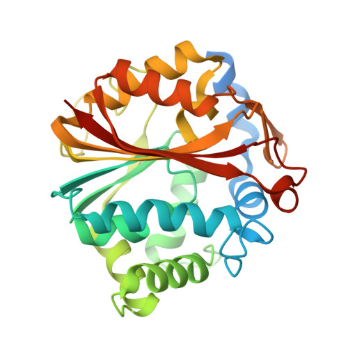
7TWU, 7TX2 - PubMed Abstract:
Phenylethanolamine N -methyltransferase (PNMT) catalyzes the S -adenosyl-l-methionine (SAM)-dependent methylation of norepinephrine to form epinephrine. Epinephrine is implicated in the regulation of blood pressure, respiration, Alzheimer's disease, and post-traumatic stress disorder (PTSD). Transition-state (TS) analogues bind their target enzymes orders of magnitude more tightly than their substrates. A synthetic strategy for first-generation TS analogues of human PNMT (hPNMT) permitted structural analysis of hPNMT and revealed potential for second-generation inhibitors [Mahmoodi, N.; J. Am. Chem. Soc. 2020, 142, 14222-14233]. A second-generation TS analogue inhibitor of PNMT was designed, synthesized, and characterized to yield a K i value of 1.2 nM. PNMT isothermal titration calorimetry (ITC) measurements of inhibitor 4 indicated a negative cooperative binding mechanism driven by large favorable entropic contributions and smaller enthalpic contributions. Cell-based assays with HEK293T cells expressing PNMT revealed a cell permeable, intracellular PNMT inhibitor with an IC 50 value of 81 nM. Structural analysis demonstrated inhibitor 4 filling catalytic site regions to recapitulate both norepinephrine and SAM interactions. Conformation of the second-generation inhibitor in the catalytic site of PNMT improves contacts relative to those from the first-generation inhibitors. Inhibitor 4 demonstrates up to 51,000-fold specificity for PNMT relative to DNA and protein methyltransferases. Inhibitor 4 also exhibits a 12,000-fold specificity for PNMT over the α 2 -adrenoceptor.
- Department of Biochemistry, Albert Einstein College of Medicine, Bronx, New York 10461, United States.
Organizational Affiliation: