HIV-2 Immature Particle Morphology Provides Insights into Gag Lattice Stability and Virus Maturation.
Talledge, N., Yang, H., Shi, K., Coray, R., Yu, G., Arndt, W.G., Meng, S., Baxter, G.C., Mendonca, L.M., Castano-Diez, D., Aihara, H., Mansky, L.M., Zhang, W.(2023) J Mol Biology 435: 168143-168143
- PubMed: 37150290 Search on PubMedSearch on PubMed Central
- DOI: https://doi.org/10.1016/j.jmb.2023.168143
- Primary Citation Related Structures:
7TV2, 8FZC - PubMed Abstract:
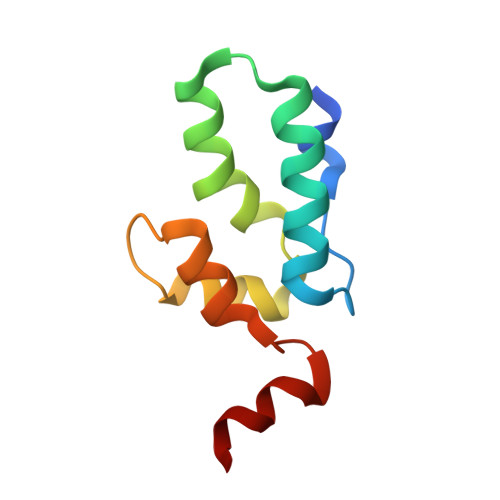
Retrovirus immature particle morphology consists of a membrane enclosed, pleomorphic, spherical and incomplete lattice of Gag hexamers. Previously, we demonstrated that human immunodeficiency virus type 2 (HIV-2) immature particles possess a distinct and extensive Gag lattice morphology. To better understand the nature of the continuously curved hexagonal Gag lattice, we have used the single particle cryo-electron microscopy method to determine the HIV-2 Gag lattice structure for immature virions. The reconstruction map at 5.5 Å resolution revealed a stable, wineglass-shaped Gag hexamer structure with structural features consistent with other lentiviral immature Gag lattice structures. Cryo-electron tomography provided evidence for nearly complete ordered Gag lattice structures in HIV-2 immature particles. We also solved a 1.98 Å resolution crystal structure of the carboxyl-terminal domain (CTD) of the HIV-2 capsid (CA) protein that identified a structured helix 12 supported via an interaction of helix 10 in the absence of the SP1 region of Gag. Residues at the helix 10-12 interface proved critical in maintaining HIV-2 particle release and infectivity. Taken together, our findings provide the first 3D organization of HIV-2 immature Gag lattice and important insights into both HIV Gag lattice stabilization and virus maturation.
- Institute for Molecular Virology, University of Minnesota - Twin Cities, Minneapolis, MN 55455, USA; Department of Diagnostic and Biological Sciences, School of Dentistry, University of Minnesota - Twin Cities, Minneapolis, MN 55455, USA; Masonic Cancer Center, University of Minnesota - Twin Cities, Minneapolis, MN 55455, USA. Electronic address: https://twitter.com/BioChemTalledge.
Organizational Affiliation: