Inhibition and Mechanism of Plasmodium falciparum Hypoxanthine-Guanine-Xanthine Phosphoribosyltransferase.
V T Minnow, Y., Suthagar, K., Clinch, K., Ducati, R.G., Ghosh, A., Buckler, J.N., Harijan, R.K., Cahill, S.M., Tyler, P.C., Schramm, V.L.(2022) ACS Chem Biol 17: 3407-3419
- PubMed: 36413975 Search on PubMedSearch on PubMed Central
- DOI: https://doi.org/10.1021/acschembio.2c00546
- Primary Citation Related Structures:
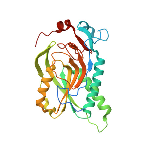
7TUX - PubMed Abstract:
Plasmodium falciparum hypoxanthine-guanine-xanthine phosphoribosyltransferase ( Pf HGXPRT) is essential for purine salvage of hypoxanthine into parasite purine nucleotides. Transition state analogue inhibitors of Pf HGXPRT are characterized by kinetic analysis, thermodynamic parameters, and X-ray crystal structures. Compound 1 , 9-deazaguanine linked to an acyclic ribocation phosphonate mimic, shows a kinetic K i of 0.5 nM. Isothermal titration calorimetry (ITC) experiments of 1 binding to Pf HGXPRT reveal enthalpically driven binding with negative cooperativity for the binding of two inhibitor molecules in the tetrameric enzyme. Crystal structures of 1 bound to Pf HGXPRT define the hydrogen bond and ionic contacts to complement binding thermodynamics. Dynamics of ribosyl transfer from 5-phospho-α-d-ribosyl 1-pyrophosphate (PRPP) to hypoxanthine were examined by 18 O isotope exchange at the bridging phosphoryl oxygen of PRPP pyrophosphate. Rotational constraints or short transition state lifetimes prevent torsional rotation and positional isotope exchange of bridging to nonbridging oxygen in the α-pyrophosphoryl group. Thermodynamic analysis of the transition state analogue and magnesium pyrophosphate binding reveal random and cooperative binding to Pf HGXPRT, unlike the obligatory ordered reaction kinetics reported earlier for substrate kinetics.
- Department of Biochemistry, Albert Einstein College of Medicine, Bronx, New York 10461, United States.
Organizational Affiliation: