7TUJ | pdb_00007tuj

NMR solution structure of the phosphorylated MUS81-binding region from human SLX4


Experimental Data Snapshot

  • Method: SOLUTION NMR
  • Conformers Calculated: 100 
  • Conformers Submitted: 20 
  • Selection Criteria: structures with the lowest energy 

wwPDB Validation 3D Report Full Report

Validation slider image for 7TUJ

This is version 1.2 of the entry. See complete history

Literature

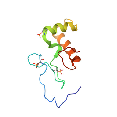
Phosphorylation of the DNA repair scaffold SLX4 drives folding of the SAP domain and activation of the MUS81-EME1 endonuclease.

Payliss, B.J.Tse, Y.W.E.Reichheld, S.E.Lemak, A.Yun, H.Y.Houliston, S.Patel, A.Arrowsmith, C.H.Sharpe, S.Wyatt, H.D.M.

(2022) Cell Rep 41: 111537-111537

  • DOI: https://doi.org/10.1016/j.celrep.2022.111537
  • Primary Citation Related Structures: 
    7TUJ

  • PubMed Abstract: 

    The DNA repair scaffold SLX4 has multifaceted roles in genome stability, many of which depend on structure-selective endonucleases. SLX4 coordinates the cell cycle-regulated assembly of SLX1, MUS81-EME1, and XPF-ERCC1 into a tri-nuclease complex called SMX. Mechanistically, how the mitotic kinase CDK1 regulates the interaction between SLX4 and MUS81-EME1 remains unclear. Here, we show that CDK1-cyclin B phosphorylates SLX4 residues T1544, T1561, and T1571 in the MUS81-binding region (SLX4 MBR ). Phosphorylated SLX4 MBR relaxes the substrate specificity of MUS81-EME1 and stimulates cleavage of replication and recombination structures, providing a biochemical explanation for the chromosome pulverization that occurs when SLX4 binds MUS81 in S-phase. Remarkably, phosphorylation of SLX4 MBR drives folding of an SAP domain, which underpins the high-affinity interaction with MUS81. We also report the structure of phosphorylated SLX4 MBR and identify the MUS81-binding interface. Our work provides mechanistic insights into how cell cycle-regulated phosphorylation of SLX4 drives the recruitment and activation of MUS81-EME1.


  • Organizational Affiliation
    • Department of Biochemistry, University of Toronto, Toronto, ON M56 1A8, Canada.

Macromolecule Content 

  • Total Structure Weight: 9.96 kDa 
  • Atom Count: 692 
  • Modeled Residue Count: 86 
  • Deposited Residue Count: 86 
  • Unique protein chains: 1

Macromolecules

Find similar proteins by:|  3D Structure
Entity ID: 1
MoleculeChains  Sequence LengthOrganismDetailsImage
Structure-specific endonuclease subunit SLX486Homo sapiensMutation(s): 0 
Gene Names: SLX4BTBD12KIAA1784KIAA1987
UniProt & NIH Common Fund Data Resources
Find proteins for Q8IY92 (Homo sapiens)
Explore Q8IY92 
Go to UniProtKB:  Q8IY92
PHAROS:  Q8IY92
GTEx:  ENSG00000188827 
Entity Groups
Sequence Clusters30% Identity50% Identity70% Identity90% Identity95% Identity100% Identity
UniProt GroupQ8IY92
Sequence Annotations
Expand
Reference Sequence

Small Molecules

Modified Residues  1 Unique
IDChains TypeFormula2D DiagramParent
TPO
Query on TPO
A
L-PEPTIDE LINKINGC4 H10 N O6 PTHR

Experimental Data & Validation

Experimental Data

  • Method: SOLUTION NMR
  • Conformers Calculated: 100 
  • Conformers Submitted: 20 
  • Selection Criteria: structures with the lowest energy 

Structure Validation

View Full Validation Report


Entry History 

& Funding Information

Deposition Data


Funding OrganizationLocationGrant Number
Canadian Institutes of Health Research (CIHR)Canada156297

Revision History  (Full details and data files)

  • Version 1.0: 2022-10-19
    Type: Initial release
  • Version 1.1: 2022-11-09
    Changes: Database references
  • Version 1.2: 2024-11-13
    Changes: Data collection, Database references, Structure summary