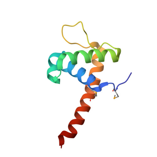
Crystal Structure of the DNA-Binding Domain of the LysR family Transcriptional Regulator YfbA from Yersinia pestis
Kim, Y., Tesar, C., Crawford, M., Chhor, G., Endres, M., Babnigg, G., Schneewind, O., Joachimiak, A., Center for Structural Genomics of Infectious Diseases (CSGID)To be published.