Elucidating Sequence and Structural Determinants of Carbohydrate Esterases for Complete Deacetylation of Substituted Xylans.
Penttinen, L., Kouhi, V., Faure, R., Skarina, T., Stogios, P., Master, E., Jurak, E.(2022) Molecules 27
- PubMed: 35566004 Search on PubMedSearch on PubMed Central
- DOI: https://doi.org/10.3390/molecules27092655
- Primary Citation Related Structures:
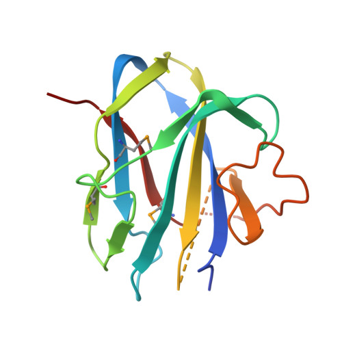
7TOG, 7TOH, 7TOI, 7TOJ, 7TOK - PubMed Abstract:
Acetylated glucuronoxylan is one of the most common types of hemicellulose in nature. The structure is formed by a β-(1→4)-linked D-xylopyranosyl (Xyl p ) backbone that can be substituted with an acetyl group at O -2 and O- 3 positions, and α-(1→2)-linked 4- O -methylglucopyranosyluronic acid (MeGlc p A). Acetyl xylan esterases (AcXE) that target mono- or doubly acetylated Xyl p are well characterized; however, the previously studied AcXE from Flavobacterium johnsoniae ( Fjo AcXE) was the first to remove the acetyl group from 2- O -MeGlc p A-3- O -acetyl-substituted Xyl p units, yet structural characteristics of these enzymes remain unspecified. Here, six homologs of Fjo AcXE were produced and three crystal structures of the enzymes were solved. Two of them are complex structures, one with bound MeGlc p A and another with acetate. All homologs were confirmed to release acetate from 2- O -MeGlc p A-3- O -acetyl-substituted xylan, and the crystal structures point to key structural elements that might serve as defining features of this unclassified carbohydrate esterase family. Enzymes comprised two domains: N-terminal CBM domain and a C-terminal SGNH domain. In Fjo AcXE and all studied homologs, the sequence motif around the catalytic serine is Gly-Asn-Ser-Ile (GNSI), which differs from other SGNH hydrolases. Binding by the MeGlc p A-Xyl p ligand is directed by positively charged and highly conserved residues at the interface of the CBM and SGNH domains of the enzyme.
- Department of Chemistry, University of Eastern Finland, 80130 Joensuu, Finland.
Organizational Affiliation: