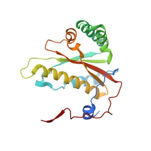
7TMG

Crystal Structure of NAD(P)H nitroreductase from Klebsiella pneumoniae (long b-axis)


Experimental Data Snapshot

  • Method: X-RAY DIFFRACTION
  • Resolution: 1.90 Å
  • R-Value Free: 0.225 
  • R-Value Work: 0.180 
  • R-Value Observed: 0.183 

wwPDB Validation   3D Report Full Report


Ligand Structure Quality Assessment 


This is version 1.1 of the entry. See complete history


Literature

Crystal Structure of NAD(P)H nitroreductase from Klebsiella pneumoniae (long b-axis)

Machen, A.J.Liu, L.Lovell, S.Battaile, K.P.Tillery, L.Shek, R.Craig, J.K.Barrett, L.K.Van Voorhis, W.C.

To be published.

Macromolecules
Find similar proteins by:  (by identity cutoff)  |  3D Structure
Entity ID: 1
MoleculeChains Sequence LengthOrganismDetailsImage
NAD(P)H nitroreductase
A, B
191Klebsiella pneumoniae subsp. pneumoniae HS11286Mutation(s): 0 
Gene Names: ydjAE3U37_20475E3U38_02825E9161_16570NCTC9504_03979
EC: 1
UniProt
Find proteins for A0A0H3GLR2 (Klebsiella pneumoniae subsp. pneumoniae (strain HS11286))
Explore A0A0H3GLR2 
Go to UniProtKB:  A0A0H3GLR2
Entity Groups  
Sequence Clusters30% Identity50% Identity70% Identity90% Identity95% Identity100% Identity
UniProt GroupA0A0H3GLR2
Sequence Annotations
Expand
  • Reference Sequence
Experimental Data & Validation

Experimental Data

Unit Cell:
Length ( Å )Angle ( ˚ )
a = 67.577α = 90
b = 99.965β = 90
c = 63.864γ = 90
Software Package:
Software NamePurpose
XDSdata reduction
Aimlessdata scaling
MOLREPphasing
PHENIXrefinement
PDB_EXTRACTdata extraction

Structure Validation

View Full Validation Report


Ligand Structure Quality Assessment 


Entry History & Funding Information

Deposition Data


Funding OrganizationLocationGrant Number
National Institutes of Health/National Institute Of Allergy and Infectious Diseases (NIH/NIAID)United StatesHHSN272201700059C

Revision History  (Full details and data files)

  • Version 1.0: 2022-02-02
    Type: Initial release
  • Version 1.1: 2023-10-18
    Changes: Data collection, Refinement description