7THZ

Structure of Leucine Rich Repeat Kinase 2's ROC domain interacting with the microtubule facing the plus end


Experimental Data Snapshot

  • Method: ELECTRON MICROSCOPY
  • Resolution: 5.00 Å
  • Aggregation State: FILAMENT 
  • Reconstruction Method: SINGLE PARTICLE 

wwPDB Validation   3D Report Full Report


This is version 1.0 of the entry. See complete history


Literature

Structural basis for Parkinson's disease-linked LRRK2's binding to microtubules.

Snead, D.M.Matyszewski, M.Dickey, A.M.Lin, Y.X.Leschziner, A.E.Reck-Peterson, S.L.

(2022) Nat Struct Mol Biol 29: 1196-1207

  • DOI: https://doi.org/10.1038/s41594-022-00863-y
  • Primary Citation of Related Structures:  
    7THY, 7THZ

  • PubMed Abstract: 

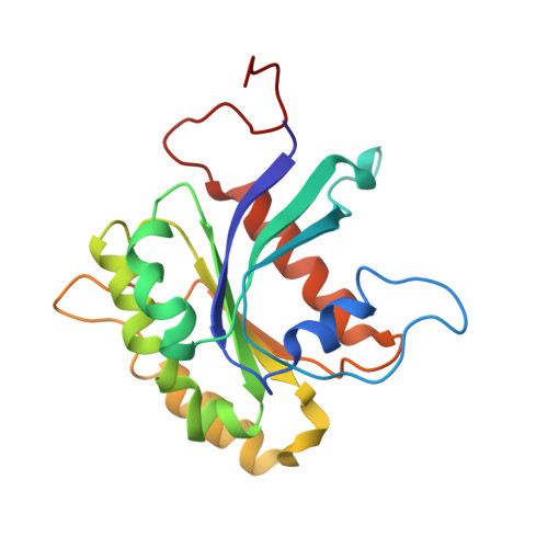
    Leucine-rich repeat kinase 2 (LRRK2) is one of the most commonly mutated genes in familial Parkinson's disease (PD). Under some circumstances, LRRK2 co-localizes with microtubules in cells, an association enhanced by PD mutations. We report a cryo-EM structure of the catalytic half of LRRK2, containing its kinase, in a closed conformation, and GTPase domains, bound to microtubules. We also report a structure of the catalytic half of LRRK1, which is closely related to LRRK2 but is not linked to PD. Although LRRK1's structure is similar to that of LRRK2, we find that LRRK1 does not interact with microtubules. Guided by these structures, we identify amino acids in LRRK2's GTPase that mediate microtubule binding; mutating them disrupts microtubule binding in vitro and in cells, without affecting LRRK2's kinase activity. Our results have implications for the design of therapeutic LRRK2 kinase inhibitors.


  • Organizational Affiliation

    Department of Cellular and Molecular Medicine, University of California, San Diego, La Jolla, CA, USA.


Macromolecules
Find similar proteins by:  (by identity cutoff)  |  3D Structure
Entity ID: 1
MoleculeChains Sequence LengthOrganismDetailsImage
Leucine-rich repeat serine/threonine-protein kinase 2194Homo sapiensMutation(s): 0 
Gene Names: LRRK2PARK8
EC: 2.7.11.1 (PDB Primary Data), 3.6.5 (PDB Primary Data)
UniProt & NIH Common Fund Data Resources
Find proteins for Q5S007 (Homo sapiens)
Explore Q5S007 
Go to UniProtKB:  Q5S007
PHAROS:  Q5S007
GTEx:  ENSG00000188906 
Entity Groups  
Sequence Clusters30% Identity50% Identity70% Identity90% Identity95% Identity100% Identity
UniProt GroupQ5S007
Sequence Annotations
Expand
  • Reference Sequence
Small Molecules
Ligands 1 Unique
IDChains Name / Formula / InChI Key2D Diagram3D Interactions
GDP
Query on GDP

Download Ideal Coordinates CCD File 
B [auth A]GUANOSINE-5'-DIPHOSPHATE
C10 H15 N5 O11 P2
QGWNDRXFNXRZMB-UUOKFMHZSA-N
Experimental Data & Validation

Experimental Data

  • Method: ELECTRON MICROSCOPY
  • Resolution: 5.00 Å
  • Aggregation State: FILAMENT 
  • Reconstruction Method: SINGLE PARTICLE 
EM Software:
TaskSoftware PackageVersion
MODEL REFINEMENTISOLDE1.2.2
MODEL REFINEMENTRosetta3.13
RECONSTRUCTIONcryoSPARC3.2

Structure Validation

View Full Validation Report


Entry History & Funding Information

Deposition Data


Funding OrganizationLocationGrant Number
Other privateUnited StatesASAP-000519
Michael J. Fox FoundationUnited States18321
National Institutes of Health/National Institute of General Medical Sciences (NIH/NIGMS)United StatesR01GM121772
National Institutes of Health/National Institute of General Medical Sciences (NIH/NIGMS)United StatesR01GM107214

Revision History  (Full details and data files)

  • Version 1.0: 2022-12-28
    Type: Initial release