7THQ

Crystal structure of PltF trapped with PigG using a proline adenosine vinylsulfonamide inhibitor


Experimental Data Snapshot

  • Method: X-RAY DIFFRACTION
  • Resolution: 2.46 Å
  • R-Value Free: 0.255 
  • R-Value Work: 0.206 
  • R-Value Observed: 0.209 

wwPDB Validation   3D Report Full Report


Ligand Structure Quality Assessment 


This is version 1.1 of the entry. See complete history


Literature

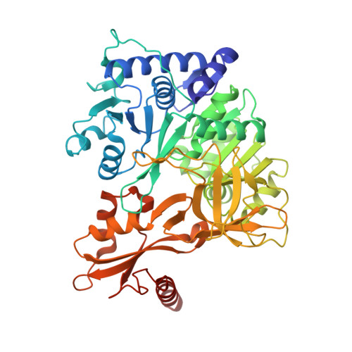
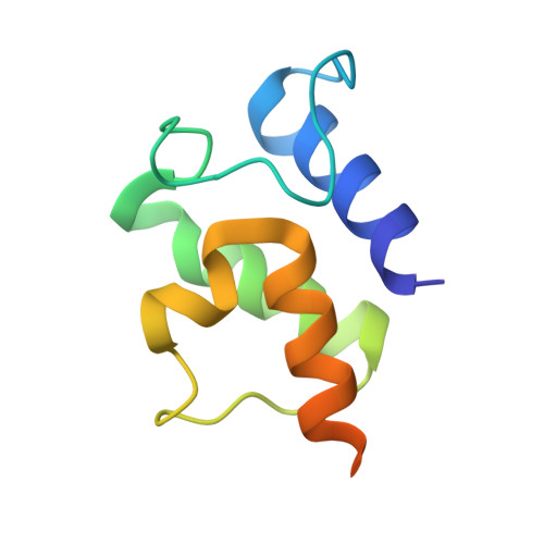
Essential Role of Loop Dynamics in Type II NRPS Biomolecular Recognition.

Corpuz, J.C.Patel, A.Davis, T.D.Podust, L.M.McCammon, J.A.Burkart, M.D.

(2022) ACS Chem Biol 17: 2890-2898

  • DOI: https://doi.org/10.1021/acschembio.2c00523
  • Primary Citation of Related Structures:  
    7THN, 7THQ

  • PubMed Abstract: 

    Non-ribosomal peptides play a critical role in the clinic as therapeutic agents. To access more chemically diverse therapeutics, non-ribosomal peptide synthetases (NRPSs) have been targeted for engineering through combinatorial biosynthesis; however, this has been met with limited success in part due to the lack of proper protein-protein interactions between non-cognate proteins. Herein, we report our use of chemical biology to enable X-ray crystallography, molecular dynamics (MD) simulations, and biochemical studies to elucidate binding specificities between peptidyl carrier proteins (PCPs) and adenylation (A) domains. Specifically, we determined X-ray crystal structures of a type II PCP crosslinked to its cognate A domain, PigG and PigI, and of PigG crosslinked to a non-cognate PigI homologue, PltF. The crosslinked PCP-A domain structures possess large protein-protein interfaces that predominantly feature hydrophobic interactions, with specific electrostatic interactions that orient the substrate for active site delivery. MD simulations of the PCP-A domain complexes and unbound PCP structures provide a dynamical evaluation of the transient interactions formed at PCP-A domain interfaces, which confirm the previously hypothesized role of a PCP loop as a crucial recognition element. Finally, we demonstrate that the interfacial interactions at the PCP loop 1 region can be modified to control PCP binding specificity through gain-of-function mutations. This work suggests that loop conformational preferences and dynamism account for improved shape complementary in the PCP-A domain interactions. Ultimately, these studies show how crystallographic, biochemical, and computational methods can be used to rationally re-engineer NRPSs for non-cognate interactions.


  • Organizational Affiliation

    Department of Chemistry and Biochemistry, University of California, San Diego, 9500 Gilman Drive, La Jolla, California 92093-0358, United States.


Macromolecules
Find similar proteins by:  (by identity cutoff)  |  3D Structure
Entity ID: 1
MoleculeChains Sequence LengthOrganismDetailsImage
L-proline--[L-prolyl-carrier protein] ligaseA [auth B],
C [auth A]
513Pseudomonas protegens Pf-5Mutation(s): 0 
Gene Names: pltFPFL_2792
EC: 6.2.1.53
UniProt
Find proteins for Q4KCY5 (Pseudomonas fluorescens (strain ATCC BAA-477 / NRRL B-23932 / Pf-5))
Explore Q4KCY5 
Go to UniProtKB:  Q4KCY5
Entity Groups  
Sequence Clusters30% Identity50% Identity70% Identity90% Identity95% Identity100% Identity
UniProt GroupQ4KCY5
Sequence Annotations
Expand
  • Reference Sequence
Find similar proteins by:  (by identity cutoff)  |  3D Structure
Entity ID: 2
MoleculeChains Sequence LengthOrganismDetailsImage
Probable acyl carrier protein PigGB [auth E],
D [auth C]
95Prodigiosinella confusarubidaMutation(s): 0 
Gene Names: pigG
UniProt
Find proteins for Q5W265 (Serratia sp. (strain ATCC 39006))
Explore Q5W265 
Go to UniProtKB:  Q5W265
Entity Groups  
Sequence Clusters30% Identity50% Identity70% Identity90% Identity95% Identity100% Identity
UniProt GroupQ5W265
Sequence Annotations
Expand
  • Reference Sequence
Small Molecules
Ligands 2 Unique
IDChains Name / Formula / InChI Key2D Diagram3D Interactions
I5M (Subject of Investigation/LOI)
Query on I5M

Download Ideal Coordinates CCD File 
H [auth E],
K [auth C]
5'-deoxy-5'-({(2S)-2-({2-[(N-{(2R)-4-[(dioxo-lambda~5~-phosphanyl)oxy]-2-hydroxy-3,3-dimethylbutanoyl}-beta-alanyl)amino]ethyl}sulfanyl)-2-[(2S)-pyrrolidin-2-yl]ethanesulfonyl}amino)adenosine
C27 H44 N9 O11 P S2
UUZQPBPCNSKBOR-LCZLERPASA-N
AZI
Query on AZI

Download Ideal Coordinates CCD File 
E [auth B]
F [auth B]
G [auth B]
I [auth A]
J [auth A]
E [auth B],
F [auth B],
G [auth B],
I [auth A],
J [auth A],
L [auth C]
AZIDE ION
N3
IVRMZWNICZWHMI-UHFFFAOYSA-N
Experimental Data & Validation

Experimental Data

  • Method: X-RAY DIFFRACTION
  • Resolution: 2.46 Å
  • R-Value Free: 0.255 
  • R-Value Work: 0.206 
  • R-Value Observed: 0.209 
  • Space Group: P 21 21 21
Unit Cell:
Length ( Å )Angle ( ˚ )
a = 65.73α = 90
b = 132.68β = 90
c = 140.94γ = 90
Software Package:
Software NamePurpose
PHENIXrefinement
PDB_EXTRACTdata extraction
XDSdata reduction
XDSdata scaling
REFMACphasing

Structure Validation

View Full Validation Report


Ligand Structure Quality Assessment 


Entry History & Funding Information

Deposition Data


Funding OrganizationLocationGrant Number
National Institutes of Health/National Institute of General Medical Sciences (NIH/NIGMS)United StatesT32 GM008326
National Institutes of Health/National Institute of General Medical Sciences (NIH/NIGMS)United States1F31GM13761601A1
National Institutes of Health/National Human Genome Research Institute (NIH/NHGRI)United StatesR01 GM095970

Revision History  (Full details and data files)

  • Version 1.0: 2022-11-30
    Type: Initial release
  • Version 1.1: 2023-10-25
    Changes: Data collection, Refinement description