7THB | pdb_00007thb

Crystal structure of an RNA-5'/DNA-3' strand exchange junction


Experimental Data Snapshot

  • Method: X-RAY DIFFRACTION
  • Resolution: 1.64 Å
  • R-Value Free: 
    0.284 (Depositor), 0.284 (DCC) 
  • R-Value Work: 
    0.237 (Depositor), 0.237 (DCC) 
  • R-Value Observed: 
    0.239 (Depositor) 

Starting Model: in silico
View more details

wwPDB Validation 3D Report Full Report

Validation slider image for 7THB

This is version 1.2 of the entry. See complete history

Literature

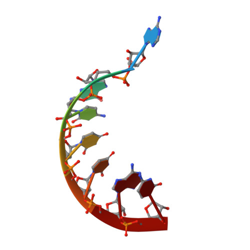
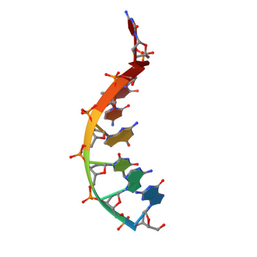
Crystal structure of an RNA/DNA strand exchange junction.

Cofsky, J.C.Knott, G.J.Gee, C.L.Doudna, J.A.

(2022) PLoS One 17: e0263547-e0263547

  • DOI: https://doi.org/10.1371/journal.pone.0263547
  • Primary Citation Related Structures: 
    7THB

  • PubMed Abstract: 

    Short segments of RNA displace one strand of a DNA duplex during diverse processes including transcription and CRISPR-mediated immunity and genome editing. These strand exchange events involve the intersection of two geometrically distinct helix types-an RNA:DNA hybrid (A-form) and a DNA:DNA homoduplex (B-form). Although previous evidence suggests that these two helices can stack on each other, it is unknown what local geometric adjustments could enable A-on-B stacking. Here we report the X-ray crystal structure of an RNA-5'/DNA-3' strand exchange junction at an anisotropic resolution of 1.6 to 2.2 Å. The structure reveals that the A-to-B helical transition involves a combination of helical axis misalignment, helical axis tilting and compression of the DNA strand within the RNA:DNA helix, where nucleotides exhibit a mixture of A- and B-form geometry. These structural principles explain previous observations of conformational stability in RNA/DNA exchange junctions, enabling a nucleic acid architecture that is repeatedly populated during biological strand exchange events.


  • Organizational Affiliation
    • Department of Molecular and Cell Biology, University of California, Berkeley, Berkeley, California, United States of America.

Macromolecule Content 

  • Total Structure Weight: 23.9 kDa 
  • Atom Count: 1,651 
  • Modeled Residue Count: 78 
  • Deposited Residue Count: 78 
  • Unique nucleic acid chains: 3

Macromolecules

Find similar nucleic acids by:  Sequence
Entity ID: 1
MoleculeChains LengthOrganismImage
DNA (5'-D(*GP*TP*AP*AP*GP*CP*AP*GP*CP*AP*TP*C)-3')
A, D, G
12synthetic construct
Sequence Annotations
Expand
Reference Sequence
Find similar nucleic acids by:  Sequence
Entity ID: 2
MoleculeChains LengthOrganismImage
RNA (5'-R(*AP*GP*CP*UP*UP*AP*C)-3')
B, E, H
7synthetic construct
Sequence Annotations
Expand
Reference Sequence
Find similar nucleic acids by:  Sequence
Entity ID: 3
MoleculeChains LengthOrganismImage
DNA (5'-D(*GP*AP*TP*GP*CP*TP*C)-3')
C, F, I
7synthetic construct
Sequence Annotations
Expand
Reference Sequence

Experimental Data & Validation

Experimental Data

  • Method: X-RAY DIFFRACTION
  • Resolution: 1.64 Å
  • R-Value Free:  0.284 (Depositor), 0.284 (DCC) 
  • R-Value Work:  0.237 (Depositor), 0.237 (DCC) 
  • R-Value Observed: 0.239 (Depositor) 
Space Group: P 1
Unit Cell:
Length ( Å )Angle ( ˚ )
a = 37.025α = 92.056
b = 43.586β = 103.722
c = 52.182γ = 99.952
Software Package:
Software NamePurpose
XDSdata reduction
STARANISOdata scaling
PHASERphasing
Cootmodel building
PHENIXrefinement

Structure Validation

View Full Validation Report


Entry History 

& Funding Information

Deposition Data


Funding OrganizationLocationGrant Number
National Science Foundation (NSF, United States)United States1817593
Howard Hughes Medical Institute (HHMI)United States--

Revision History  (Full details and data files)

  • Version 1.0: 2022-04-27
    Type: Initial release
  • Version 1.1: 2024-02-28
    Changes: Data collection
  • Version 1.2: 2024-04-03
    Changes: Refinement description