A comparison of the bacterial CYP51 cytochrome P450 enzymes from Mycobacterium marinum and Mycobacterium tuberculosis.
Mohamed, H., Child, S.A., Bruning, J.B., Bell, S.G.(2022) J Steroid Biochem Mol Biol 221: 106097-106097
- PubMed: 35346833 Search on PubMed
- DOI: https://doi.org/10.1016/j.jsbmb.2022.106097
- Primary Citation Related Structures:
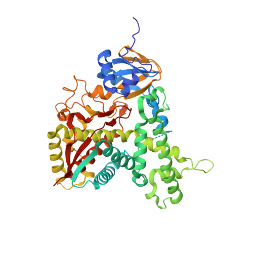
7TEF - PubMed Abstract:
Members of the CYP51 family of cytochrome P450 enzymes are classified as sterol demethylases involved in the metabolic formation of cholesterol and related derivatives. The CYP51 enzyme from Mycobacterium marinum was studied and compared to its counterpart from Mycobacterium tuberculosis to determine the degree of functional conservation between them. Spectroscopic analyses of substrate and inhibitor binding of the purified CYP51 enzymes from M. marinum and M. tuberculosis were performed. The catalytic oxidation of lanosterol and related steroids was investigated. M. marinum CYP51 was structurally characterized by X-ray crystallography. The CYP51 enzyme of M. marinum is sequentially closely related to CYP51B1 from M. tuberculosis. However, differences in the heme spin state of each enzyme were observed upon the addition of steroids and other ligands. Both enzymes displayed different binding properties to those reported for the CYP51-Fdx fusion protein from the bacterium Methylococcus capsulatus. The enzymes were able to oxidatively demethylate lanosterol to generate 14-demethylanosterol, but no products were detected for the related species dihydrolanosterol and eburicol. The crystal structure of CYP51 from M. marinum in the absence of added substrate but with a Bis-Tris molecule within the active site was resolved. The CYP51 enzyme of M. marinum displays differences in how steroids and other ligands bind compared to the M. tuberculosis enzyme. This was related to structural differences between the two enzymes. Overall, both of these CYP51 enzymes from mycobacterial species displayed significant differences to the CYP51 enzymes of eukaryotic species and the bacterial CYP51-Fdx enzyme of Me. capsulatus.
- Department of Chemistry, University of Adelaide, SA 5005, Australia.
Organizational Affiliation: