Determination of the pH dependence, substrate specificity, and turnovers of alternative substrates for human ornithine aminotransferase.
Butrin, A., Butrin, A., Wawrzak, Z., Moran, G.R., Liu, D.(2022) J Biol Chem 298: 101969-101969
- PubMed: 35460691 Search on PubMedSearch on PubMed Central
- DOI: https://doi.org/10.1016/j.jbc.2022.101969
- Primary Citation Related Structures:
7T9Z, 7TA0, 7TA1 - PubMed Abstract:
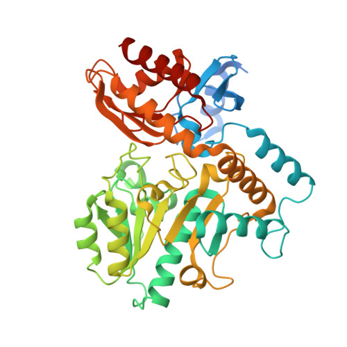
Hepatocellular carcinoma (HCC) is the most common primary cancer of the liver and occurs predominantly in patients with underlying chronic liver diseases. Over the past decade, human ornithine aminotransferase (hOAT), which is an enzyme that catalyzes the metabolic conversion of ornithine into an intermediate for proline or glutamate synthesis, has been found to be overexpressed in HCC cells. hOAT has since emerged as a promising target for novel anticancer therapies, especially for the ongoing rational design effort to discover mechanism-based inactivators (MBIs). Despite the significance of hOAT in human metabolism and its clinical potential as a drug target against HCC, there are significant knowledge deficits with regard to its catalytic mechanism and structural characteristics. Ongoing MBI design efforts require in-depth knowledge of the enzyme active site, in particular, pKa values of potential nucleophiles and residues necessary for the molecular recognition of ligands. Here, we conducted a study detailing the fundamental active-site properties of hOAT using stopped-flow spectrophotometry and X-ray crystallography. Our results quantitatively revealed the pH dependence of the multistep reaction mechanism and illuminated the roles of ornithine α-amino and δ-amino groups in substrate recognition and in facilitating catalytic turnover. These findings provided insights of the catalytic mechanism that could benefit the rational design of MBIs against hOAT. In addition, substrate recognition and turnover of several fragment-sized alternative substrates of hOATs, which could serve as structural templates for MBI design, were also elucidated.
- Department of Chemistry and Biochemistry, Loyola University Chicago, Chicago, Illinois, USA.
Organizational Affiliation: