


Structure Characterization of Zinc Finger Motif 1 and 2 of GLI1 DNA Binding Region
Wu, M., Jahan, N., Sharp, A., Ullah, A., Augelli-Szafran, C.E., Zhang, S., Boohaker, R.J.(2024) Int J Mol Sci 25
Experimental Data Snapshot
Starting Model: experimental
View more details
wwPDB Validation 3D Report Full Report

(2024) Int J Mol Sci 25
Entity ID: 1 | |||||
|---|---|---|---|---|---|
| Molecule | Chains | Sequence Length | Organism | Details | Image |
| Isoform 2 of Zinc finger protein GLI1 | 71 | Homo sapiens | Mutation(s): 0 Gene Names: GLI1, GLI | 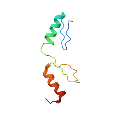 | |
UniProt & NIH Common Fund Data Resources | |||||
GTEx: ENSG00000111087 | |||||
Entity Groups | |||||
| Sequence Clusters | 30% Identity50% Identity70% Identity90% Identity95% Identity100% Identity | ||||
| UniProt Group | P08151 | ||||
Sequence AnnotationsExpand | |||||
Reference Sequence | |||||
| Ligands 1 Unique | |||||
|---|---|---|---|---|---|
| ID | Chains | Name / Formula / InChI Key | 2D Diagram | 3D Interactions | |
| ZN (Subject of Investigation/LOI) Download:Ideal Coordinates CCD File | C [auth A], D [auth A], E [auth B], F [auth B] | ZINC ION Zn PTFCDOFLOPIGGS-UHFFFAOYSA-N |  | ||
| Length ( Å ) | Angle ( ˚ ) |
|---|---|
| a = 66.728 | α = 90 |
| b = 66.728 | β = 90 |
| c = 65.909 | γ = 120 |
| Software Name | Purpose |
|---|---|
| XDS | data reduction |
| SCALA | data scaling |
| PHASER | phasing |
| REFMAC | refinement |
| PDB_EXTRACT | data extraction |
| Funding Organization | Location | Grant Number |
|---|---|---|
| National Institutes of Health/National Cancer Institute (NIH/NCI) | United States | 5RO1CA183921-05 |


