7T8S | pdb_00007t8s

Light Harvesting complex phycoerythrin PE 566, from the cryptophyte Cryptomonas pyrenoidifera


Experimental Data Snapshot

  • Method: X-RAY DIFFRACTION
  • Resolution: 2.00 Å
  • R-Value Free: 
    0.234 (Depositor), 0.235 (DCC) 
  • R-Value Work: 
    0.203 (Depositor), 0.203 (DCC) 
  • R-Value Observed: 
    0.204 (Depositor) 

Starting Model: experimental
View more details

wwPDB Validation 3D Report Full Report

Validation slider image for 7T8S

Ligand Structure Quality Assessment 


This is version 1.2 of the entry. See complete history

Literature

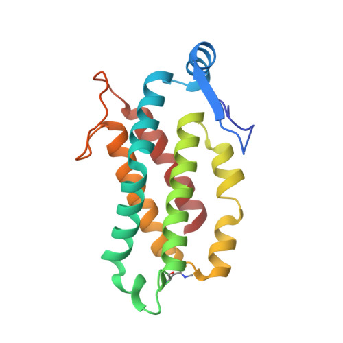
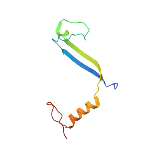
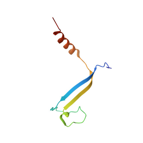
Molecular structures reveal the origin of spectral variation in cryptophyte light harvesting antenna proteins.

Michie, K.A.Harrop, S.J.Rathbone, H.W.Wilk, K.E.Teng, C.Y.Hoef-Emden, K.Hiller, R.G.Green, B.R.Curmi, P.M.G.

(2023) Protein Sci 32: e4586-e4586

  • DOI: https://doi.org/10.1002/pro.4586
  • Primary Citation Related Structures: 
    7T7U, 7T89, 7T8S

  • PubMed Abstract: 

    In addition to their membrane-bound chlorophyll a/c light-harvesting antenna, the cryptophyte algae have evolved a unique phycobiliprotein antenna system located in the thylakoid lumen. The basic unit of this antenna consists of two copies of an αβ protomer where the α and β subunits scaffold different combinations of a limited number of linear tetrapyrrole chromophores. While the β subunit is highly conserved, encoded by a single plastid gene, the nuclear-encoded α subunits have evolved diversified multigene families. It is still unclear how this sequence diversity results in the spectral diversity of the mature proteins. By careful examination of three newly determined crystal structures in comparison with three previously obtained, we show how the α subunit amino acid sequences control chromophore conformations and hence spectral properties even when the chromophores are identical. Previously we have shown that α subunits control the quaternary structure of the mature αβ.αβ complex (either open or closed), however, each species appeared to only harbor a single quaternary form. Here we show that species of the Hemiselmis genus contain expressed α subunit genes that encode both distinct quaternary structures. Finally, we have discovered a common single-copy gene (expressed into protein) consisting of tandem copies of a small α subunit that could potentially scaffold pairs of light harvesting units. Together, our results show how the diversity of the multigene α subunit family produces a range of mature cryptophyte antenna proteins with differing spectral properties, and the potential for minor forms that could contribute to acclimation to varying light regimes.


  • Organizational Affiliation
    • School of Physics, The University of New South Wales, Sydney, New South Wales, Australia.

Macromolecule Content 

  • Total Structure Weight: 232.32 kDa 
  • Atom Count: 17,083 
  • Modeled Residue Count: 1,933 
  • Deposited Residue Count: 2,016 
  • Unique protein chains: 3

Macromolecules

Find similar proteins by:|  3D Structure
Entity ID: 1
MoleculeChains  Sequence LengthOrganismDetailsImage
Phycoerythrin beta subunit
A, C, E, G, I
A, C, E, G, I, K, M, O
178Cryptomonas pyrenoidiferaMutation(s): 0 
UniProt
Find proteins for A0A222AH92 (Cryptomonas curvata)
Explore A0A222AH92 
Go to UniProtKB:  A0A222AH92
Entity Groups
Sequence Clusters30% Identity50% Identity70% Identity90% Identity95% Identity100% Identity
UniProt GroupA0A222AH92
Sequence Annotations
Expand
Reference Sequence
Find similar proteins by:|  3D Structure
Entity ID: 2
MoleculeChains  Sequence LengthOrganismDetailsImage
phycoerythrin alpha-1 subunit
B, F, J, N
78Cryptomonas pyrenoidiferaMutation(s): 0 
UniProt
Find proteins for A0A7S0TJJ1 (Hemiselmis andersenii)
Explore A0A7S0TJJ1 
Go to UniProtKB:  A0A7S0TJJ1
Entity Groups
Sequence Clusters30% Identity50% Identity70% Identity90% Identity95% Identity100% Identity
UniProt GroupA0A7S0TJJ1
Sequence Annotations
Expand
Reference Sequence
Find similar proteins by:|  3D Structure
Entity ID: 3
MoleculeChains  Sequence LengthOrganismDetailsImage
phycoerythrin alpha-2 subunit
D, H, L, P
70Cryptomonas pyrenoidiferaMutation(s): 0 
UniProt
Find proteins for A0A7S0UC19 (Hemiselmis andersenii)
Explore A0A7S0UC19 
Go to UniProtKB:  A0A7S0UC19
Entity Groups
Sequence Clusters30% Identity50% Identity70% Identity90% Identity95% Identity100% Identity
UniProt GroupA0A7S0UC19
Sequence Annotations
Expand
Reference Sequence

Small Molecules

Ligands 5 Unique
IDChains Name / Formula / InChI Key2D Diagram3D Interactions
KQ6
(Subject of Investigation/LOI)

Query on KQ6


Download:Ideal Coordinates CCD File
CA [auth E]
DB [auth O]
HA [auth G]
NA [auth I]
S [auth A]
CA [auth E],
DB [auth O],
HA [auth G],
NA [auth I],
S [auth A],
TA [auth K],
XA [auth M],
Y [auth C]
Bilin 584 (doubly linked)
C33 H40 N4 O6
KXOMFZIVZHIPRO-GUEBWASOSA-N
PEB
(Subject of Investigation/LOI)

Query on PEB


Download:Ideal Coordinates CCD File
AA [auth E]
BB [auth O]
FA [auth G]
LA [auth I]
Q [auth A]
AA [auth E],
BB [auth O],
FA [auth G],
LA [auth I],
Q [auth A],
RA [auth K],
VA [auth M],
W [auth C]
PHYCOERYTHROBILIN
C33 H40 N4 O6
NKCBCVIFPXGHAV-WAVSMFBNSA-N
KPX
(Subject of Investigation/LOI)

Query on KPX


Download:Ideal Coordinates CCD File
BA [auth E]
CB [auth O]
GA [auth G]
MA [auth I]
R [auth A]
BA [auth E],
CB [auth O],
GA [auth G],
MA [auth I],
R [auth A],
SA [auth K],
WA [auth M],
X [auth C]
Bilin 584 (single linked)
C33 H38 N4 O6
DELSVXYVYXKOJW-GUEBWASOSA-N
KP9
(Subject of Investigation/LOI)

Query on KP9


Download:Ideal Coordinates CCD File
EA [auth F]
EB [auth P]
JA [auth H]
QA [auth J]
UA [auth L]
EA [auth F],
EB [auth P],
JA [auth H],
QA [auth J],
UA [auth L],
V [auth B],
Z [auth D],
ZA [auth N]
Bilin 618 (single linked)
C33 H36 N4 O6
DWOKJMXOEBXRPB-ZHKKBVGESA-N
GOL

Query on GOL


Download:Ideal Coordinates CCD File
AB [auth N]
DA [auth E]
IA [auth G]
KA [auth H]
OA [auth I]
AB [auth N],
DA [auth E],
IA [auth G],
KA [auth H],
OA [auth I],
PA [auth I],
T [auth A],
U [auth A],
YA [auth M]
GLYCEROL
C3 H8 O3
PEDCQBHIVMGVHV-UHFFFAOYSA-N
Modified Residues  1 Unique
IDChains TypeFormula2D DiagramParent
MEN
Query on MEN
A, C, E, G, I
A, C, E, G, I, K, M, O
L-PEPTIDE LINKINGC5 H10 N2 O3ASN

Experimental Data & Validation

Experimental Data

  • Method: X-RAY DIFFRACTION
  • Resolution: 2.00 Å
  • R-Value Free:  0.234 (Depositor), 0.235 (DCC) 
  • R-Value Work:  0.203 (Depositor), 0.203 (DCC) 
  • R-Value Observed: 0.204 (Depositor) 
Space Group: P 41 21 2
Unit Cell:
Length ( Å )Angle ( ˚ )
a = 178.359α = 90
b = 178.359β = 90
c = 173.546γ = 90
Software Package:
Software NamePurpose
PHENIXrefinement
Aimlessdata scaling
PHASERphasing
MOSFLMdata reduction

Structure Validation

View Full Validation Report


Ligand Structure Quality Assessment 


Entry History 

& Funding Information

Deposition Data


Funding OrganizationLocationGrant Number
Australian Research Council (ARC)ARC LIEF LE190100165
Australian Research Council (ARC)DP180103964
Other governmentFA2386-17-1-4101

Revision History  (Full details and data files)

  • Version 1.0: 2023-02-15
    Type: Initial release
  • Version 1.1: 2023-03-08
    Changes: Database references
  • Version 1.2: 2023-10-25
    Changes: Data collection, Refinement description