7T6H | pdb_00007t6h

E. coli dihydroorotate dehydrogenase


Experimental Data Snapshot

  • Method: X-RAY DIFFRACTION
  • Resolution: 2.42 Å
  • R-Value Free: 
    0.235 (Depositor), 0.234 (DCC) 
  • R-Value Work: 
    0.200 (Depositor), 0.201 (DCC) 
  • R-Value Observed: 
    0.202 (Depositor) 

wwPDB Validation 3D Report Full Report

Validation slider image for 7T6H

Ligand Structure Quality Assessment 


This is version 1.2 of the entry. See complete history

Literature

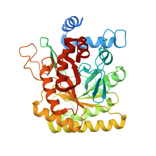
Structural insights into inhibition of the drug target dihydroorotate dehydrogenase by bacterial hydroxyalkylquinolines.

Horwitz, S.M.Blue, T.C.Ambarian, J.A.Hoshino, S.Seyedsayamdost, M.R.Davis, K.M.

(2022) RSC Chem Biol 3: 420-425

  • DOI: https://doi.org/10.1039/d1cb00255d
  • Primary Citation Related Structures: 
    7T5K, 7T5Y, 7T6C, 7T6H

  • PubMed Abstract: 

    Hydroxyalkylquinolines (HAQs) are ubiquitious natural products but their interactions with associated protein targets remain elusive. We report X-ray crystal structures of two HAQs in complex with dihydroorotate dehydrogenase (DHODH). Our results reveal the structural basis of DHODH inhibition by HAQs and open the door to downstream structure-activity relationship studies.


  • Organizational Affiliation
    • Department of Chemistry, Emory University Atlanta GA 30322 USA katherine.davis@emory.edu.

Macromolecule Content 

  • Total Structure Weight: 81.8 kDa 
  • Atom Count: 5,343 
  • Modeled Residue Count: 670 
  • Deposited Residue Count: 736 
  • Unique protein chains: 1

Macromolecules

Find similar proteins by:|  3D Structure
Entity ID: 1
MoleculeChains  Sequence LengthOrganismDetailsImage
Dihydroorotate dehydrogenase (quinone)
A, B
368Escherichia coli K-12Mutation(s): 0 
Gene Names: pyrDb0945JW0928
EC: 1.3.5.2
UniProt
Find proteins for P0A7E1 (Escherichia coli (strain K12))
Explore P0A7E1 
Go to UniProtKB:  P0A7E1
Entity Groups
Sequence Clusters30% Identity50% Identity70% Identity90% Identity95% Identity100% Identity
UniProt GroupP0A7E1
Sequence Annotations
Expand
Reference Sequence

Experimental Data & Validation

Experimental Data

  • Method: X-RAY DIFFRACTION
  • Resolution: 2.42 Å
  • R-Value Free:  0.235 (Depositor), 0.234 (DCC) 
  • R-Value Work:  0.200 (Depositor), 0.201 (DCC) 
  • R-Value Observed: 0.202 (Depositor) 
Space Group: C 2 2 21
Unit Cell:
Length ( Å )Angle ( ˚ )
a = 104.525α = 90
b = 169.495β = 90
c = 129.984γ = 90
Software Package:
Software NamePurpose
PHENIXrefinement
Cootmodel building
XDSdata reduction
XDSdata scaling
PHASERphasing

Structure Validation

View Full Validation Report


Ligand Structure Quality Assessment 


Entry History 

& Funding Information

Deposition Data


Funding OrganizationLocationGrant Number
National Institutes of Health/National Institute of General Medical Sciences (NIH/NIGMS)United States--

Revision History  (Full details and data files)

  • Version 1.0: 2022-03-02
    Type: Initial release
  • Version 1.1: 2022-06-29
    Changes: Database references
  • Version 1.2: 2024-02-28
    Changes: Data collection