Molecular Basis of Functional Effects of Phosphorylation of the C-Terminal Domain of the Rabies Virus P Protein.
Zhan, J., Watts, E., Brice, A.M., Metcalfe, R.D., Rozario, A.M., Sethi, A., Yan, F., Bell, T.D.M., Griffin, M.D.W., Moseley, G.W., Gooley, P.R.(2022) J Virol 96: e0011122-e0011122
- PubMed: 35404083 Search on PubMedSearch on PubMed Central
- DOI: https://doi.org/10.1128/jvi.00111-22
- Primary Citation Related Structures:
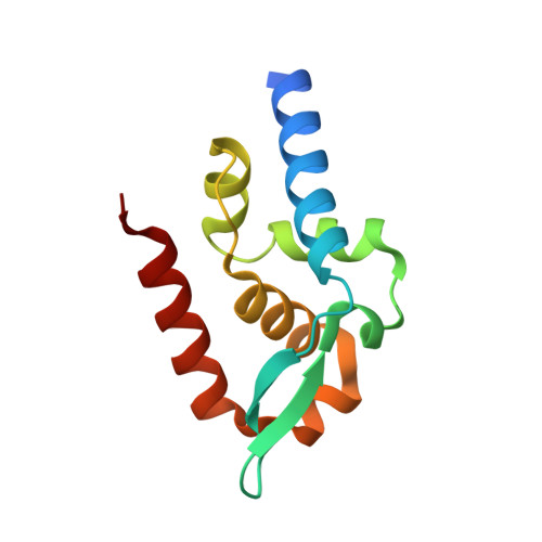
7T5G, 7T5H - PubMed Abstract:
The rabies virus (RABV) phosphoprotein (P protein) is expressed as several isoforms, which differ in nucleocytoplasmic localization and microtubule (MT) association, mediated by several sequences, including nuclear localization (NLS) and export (NES) sequences. This appears to underpin a functional diversity enabling multiple functions in viral replication and modulation of host biology. Mechanisms regulating trafficking are poorly defined, but phosphorylation by protein kinase C (PKC) in the P protein C-terminal domain (P CTD ) regulates nuclear trafficking, mediated by P CTD -localized NLS/NES sequences, indicating that phosphorylation contributes to functional diversity. The molecular mechanism underlying the effects of PKC, and potential roles in regulating other host-cell interactions are unresolved. Here, we assess effects of phosphorylation on the P3 isoform, which differs from longer isoforms through an ability to localize to the nucleus and associate with MTs, which are associated with antagonism of interferon (IFN) signaling. We find that phosphomimetic mutation of the PKC site S210 inhibits nuclear accumulation and MT association/bundling. Structural analysis indicated that phosphomimetic mutation induces no significant structural change to the NLS/NES but results in the side chain of N226 switching its interactions from E228, within the NES, to E210. Intriguingly, N226 is the sole substituted residue between the P CTD of the pathogenic IFN-resistant RABV strain Nishigahara and a derivative attenuated IFN-sensitive strain Ni-CE, inhibiting P3 nuclear localization and MT association. Thus, S210 phosphorylation appears to impact on N226/E228 to regulate P protein localization, with N226 mutation in Ni-CE mimicking a constitutively phosphorylated state resulting in IFN sensitivity and attenuation. IMPORTANCE Rabies virus P protein is a multifunctional protein with critical roles in replication and manipulation of host-cell processes, including subversion of immunity. This functional diversity involves interactions of several P protein isoforms with the cell nucleus and microtubules. Previous studies showed that phosphorylation of the P protein C-terminal domain (P CTD ) at S210, near nuclear trafficking sequences, regulates nucleocytoplasmic localization, indicating key roles in functional diversity. The molecular mechanisms of this regulation have remained unknown. Here, we show that phosphomimetic mutation of S210 regulates nuclear localization and MT association. This regulation does not appear to result from disrupted P CTD structure, but rather from a switch of specific side chain interactions of N226. Intriguingly, N226 was previously implicated in P protein nuclear localization/MT association, immune evasion, and RABV pathogenesis, through undefined mechanisms. Our data indicate that the S210-N226 interface is a key regulator of virus-host interactions, which is significant for pathogenesis.
- Department of Biochemistry and Pharmacology, Bio21 Molecular Science and Biotechnology Institute, University of Melbournegrid.1008.9, Parkville, Victoria, Australia.
Organizational Affiliation: