Funding Organization(s): National Institutes of Health/National Institute Of Allergy and Infectious Diseases (NIH/NIAID), Bill & Melinda Gates Foundation
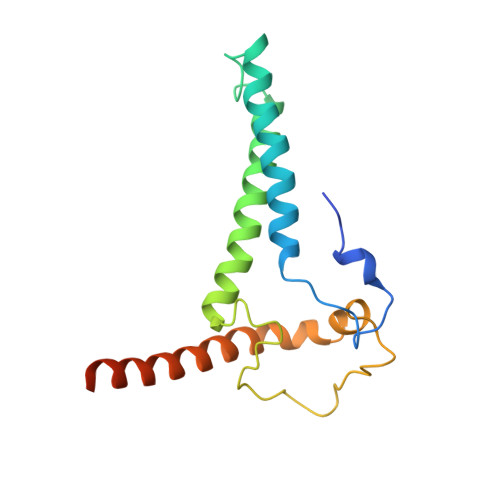
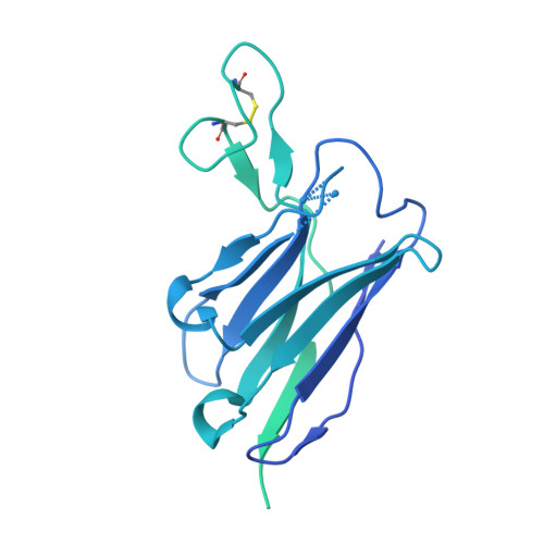
SIVmac239 infection of macaques is a favored model of human HIV infection. However, the SIVmac239 envelope (Env) trimer structure, glycan occupancy, and the targets and ability of neutralizing antibodies (nAbs) to protect against SIVmac239 remain unknown. Here, we report the isolation of SIVmac239 nAbs that recognize a glycan hole and the V1/V4 loop. A high-resolution structure of a SIVmac239 Env trimer-nAb complex shows many similarities to HIV and SIVcpz Envs, but with distinct V4 features and an extended V1 loop. Moreover, SIVmac239 Env has a higher glycan shield density than HIV Env that may contribute to poor or delayed nAb responses in SIVmac239-infected macaques. Passive transfer of a nAb protects macaques from repeated intravenous SIVmac239 challenge at serum titers comparable to those described for protection of humans against HIV infection. Our results provide structural insights for vaccine design and shed light on antibody-mediated protection in the SIV model.
Organizational Affiliation:
Department of Immunology and Microbiology, The Scripps Research Institute, La Jolla, CA, 92037, USA.
IAVI Neutralizing Antibody Center, The Scripps Research Institute, La Jolla, CA, 92037, USA.
Consortium for HIV/AIDS Vaccine Development (CHAVD), The Scripps Research Institute, La Jolla, CA, 92037, USA.
Department of Integrative Structural and Computational Biology, The Scripps Research Institute, La Jolla, CA, 92037, USA.
Department of Pathology, George Washington University, Washington, DC, 20037, USA.
School of Biological Sciences, University of Southampton, Southampton, SO17 1BJ, UK.
Theoretical Biology and Biophysics Group, Los Alamos National Laboratory, Los Alamos, NM, 87545, USA.
IAVI, New York, NY, 10004, USA.
Wisconsin National Primate Research Center, University of Wisconsin-Madison, Madison, WI, 53715, USA.
AIDS and Cancer Virus Program, Frederick National Laboratory for Cancer Research, Frederick, MD, 21701, USA.
Department of Pathology, Miller School of Medicine, University of Miami, Miami, FL, 33136, USA.
Department of Immunology and Microbiology, The Scripps Research Institute, La Jolla, CA, 92037, USA. andrabi@scripps.edu.
IAVI Neutralizing Antibody Center, The Scripps Research Institute, La Jolla, CA, 92037, USA. andrabi@scripps.edu.
Consortium for HIV/AIDS Vaccine Development (CHAVD), The Scripps Research Institute, La Jolla, CA, 92037, USA. andrabi@scripps.edu.
IAVI Neutralizing Antibody Center, The Scripps Research Institute, La Jolla, CA, 92037, USA. andrew@scripps.edu.
Consortium for HIV/AIDS Vaccine Development (CHAVD), The Scripps Research Institute, La Jolla, CA, 92037, USA. andrew@scripps.edu.
Department of Integrative Structural and Computational Biology, The Scripps Research Institute, La Jolla, CA, 92037, USA. andrew@scripps.edu.
Department of Immunology and Microbiology, The Scripps Research Institute, La Jolla, CA, 92037, USA. burton@scripps.edu.
IAVI Neutralizing Antibody Center, The Scripps Research Institute, La Jolla, CA, 92037, USA. burton@scripps.edu.
Consortium for HIV/AIDS Vaccine Development (CHAVD), The Scripps Research Institute, La Jolla, CA, 92037, USA. burton@scripps.edu.
Ragon Institute of Massachusetts General Hospital, Massachusetts Institute of Technology, and Harvard University, Cambridge, MA, 02139, USA. burton@scripps.edu.
Department of Immunology and Microbiology, The Scripps Research Institute, La Jolla, CA, 92037, USA. dsok@iavi.org.
IAVI Neutralizing Antibody Center, The Scripps Research Institute, La Jolla, CA, 92037, USA. dsok@iavi.org.
Consortium for HIV/AIDS Vaccine Development (CHAVD), The Scripps Research Institute, La Jolla, CA, 92037, USA. dsok@iavi.org.
AA [auth B] BA [auth B] CA [auth B] Q [auth A] R [auth A]
AA [auth B], BA [auth B], CA [auth B], Q [auth A], R [auth A], S [auth A], T [auth A], U [auth A], V [auth A], W [auth A], X [auth A], Y [auth A], Z [auth A]
2-acetamido-2-deoxy-beta-D-glucopyranose C8 H15 N O6 OVRNDRQMDRJTHS-FMDGEEDCSA-N