7SYB | pdb_00007syb

Crystal Structure of sulfurtransferase (DsrC family protein) from Acinetobacter baumannii


Experimental Data Snapshot

  • Method: X-RAY DIFFRACTION
  • Resolution: 1.45 Å
  • R-Value Free: 
    0.168 (Depositor), 0.186 (DCC) 
  • R-Value Work: 
    0.139 (Depositor) 
  • R-Value Observed: 
    0.142 (Depositor) 

wwPDB Validation 3D Report Full Report

Validation slider image for 7SYB

This is version 1.1 of the entry. See complete history

Literature

Crystal Structure of sulfurtransferase (DsrC family protein) from Acinetobacter baumannii

Calhoun, B.M.Bolejack, M.J.Lorimer, D.D.Horanyi, P.S.Edwards, T.E.

To be published.

Macromolecule Content 

  • Total Structure Weight: 12.74 kDa 
  • Atom Count: 1,031 
  • Modeled Residue Count: 103 
  • Deposited Residue Count: 111 
  • Unique protein chains: 1

Macromolecules

Find similar proteins by:|  3D Structure
Entity ID: 1
MoleculeChains  Sequence LengthOrganismDetailsImage
Sulfurtransferase111Acinetobacter baumanniiMutation(s): 0 
Gene Names: tusE
EC: 2.8.1
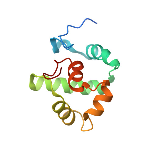
Experimental Data & Validation

Experimental Data

  • Method: X-RAY DIFFRACTION
  • Resolution: 1.45 Å
  • R-Value Free:  0.168 (Depositor), 0.186 (DCC) 
  • R-Value Work:  0.139 (Depositor) 
  • R-Value Observed: 0.142 (Depositor) 
Space Group: P 1 21 1
Unit Cell:
Length ( Å )Angle ( ˚ )
a = 32.5α = 90
b = 46.84β = 108.02
c = 39.48γ = 90
Software Package:
Software NamePurpose
XDSdata reduction
XSCALEdata scaling
PHENIXrefinement
PDB_EXTRACTdata extraction
MR-Rosettaphasing

Structure Validation

View Full Validation Report


Entry History 

& Funding Information

Deposition Data


Funding OrganizationLocationGrant Number
National Institutes of Health/National Institute Of Allergy and Infectious Diseases (NIH/NIAID)United States--

Revision History  (Full details and data files)

  • Version 1.0: 2021-12-08
    Type: Initial release
  • Version 1.1: 2024-02-28
    Changes: Data collection