7STZ

Crystal Structure of Human E-cadherin EC1-5 bound by mouse monoclonal antibody Fab mAb-1_19A11


Experimental Data Snapshot

  • Method: X-RAY DIFFRACTION
  • Resolution: 2.95 Å
  • R-Value Free: 0.208 
  • R-Value Work: 0.182 
  • R-Value Observed: 0.182 

wwPDB Validation   3D Report Full Report


Ligand Structure Quality Assessment 


This is version 1.2 of the entry. See complete history


Literature

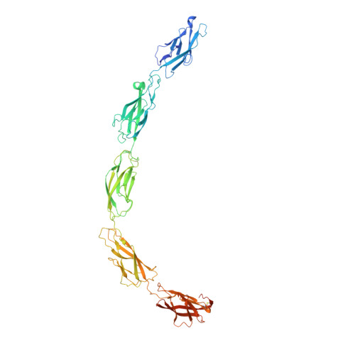
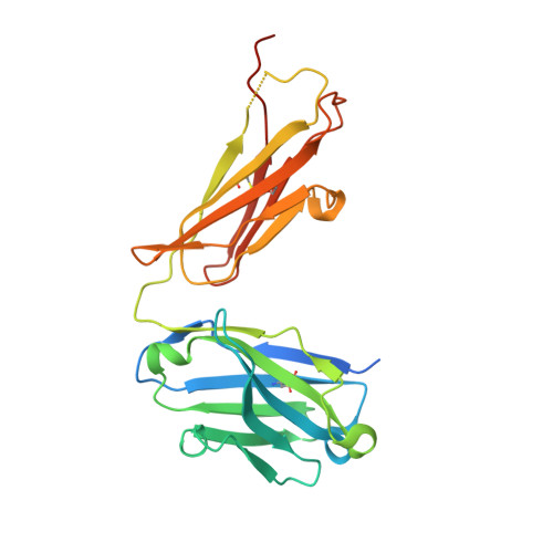
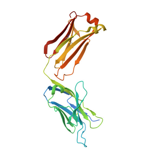
Regulation of multiple dimeric states of E-cadherin by adhesion activating antibodies revealed through Cryo-EM and X-ray crystallography.

Maker, A.Bolejack, M.Schecterson, L.Hammerson, B.Abendroth, J.Edwards, T.E.Staker, B.Myler, P.J.Gumbiner, B.M.

(2022) PNAS Nexus 1: pgac163-pgac163

  • DOI: https://doi.org/10.1093/pnasnexus/pgac163
  • Primary Citation of Related Structures:  
    6VEL, 7STZ

  • PubMed Abstract: 

    E-cadherin adhesion is regulated at the cell surface, a process that can be replicated by activating antibodies. We use cryo-electron microscopy (EM) and X-ray crystallography to examine functional states of the cadherin adhesive dimer. This dimer is mediated by N-terminal beta strand-swapping involving Trp2, and forms via a different transient X-dimer intermediate. X-dimers are observed in cryo-EM along with monomers and strand-swap dimers, indicating that X-dimers form stable interactions. A novel EC4-mediated dimer was also observed. Activating Fab binding caused no gross structural changes in E-cadherin monomers, but can facilitate strand swapping. Moreover, activating Fab binding is incompatible with the formation of the X-dimer. Both cryo-EM and X-ray crystallography reveal a distinctive twisted strand-swap dimer conformation caused by an outward shift in the N-terminal beta strand that may represent a strengthened state. Thus, regulation of adhesion involves changes in cadherin dimer configurations.


  • Organizational Affiliation

    Department of Biochemistry, University of Washington, USA.


Macromolecules
Find similar proteins by:  (by identity cutoff)  |  3D Structure
Entity ID: 1
MoleculeChains Sequence LengthOrganismDetailsImage
Cadherin-1A [auth C],
D
590Homo sapiensMutation(s): 0 
Gene Names: CDH1CDHEUVO
UniProt & NIH Common Fund Data Resources
Find proteins for P12830 (Homo sapiens)
Explore P12830 
Go to UniProtKB:  P12830
PHAROS:  P12830
GTEx:  ENSG00000039068 
Entity Groups  
Sequence Clusters30% Identity50% Identity70% Identity90% Identity95% Identity100% Identity
UniProt GroupP12830
Sequence Annotations
Expand
  • Reference Sequence
Find similar proteins by:  (by identity cutoff)  |  3D Structure
Entity ID: 2
MoleculeChains Sequence LengthOrganismDetailsImage
mAb-1_19A11 Heavy ChainB [auth H],
E [auth I]
246Mus musculusMutation(s): 0 
Gene Names: Igh
Entity Groups  
Sequence Clusters30% Identity50% Identity70% Identity90% Identity95% Identity100% Identity
Sequence Annotations
Expand
  • Reference Sequence
Find similar proteins by:  (by identity cutoff)  |  3D Structure
Entity ID: 3
MoleculeChains Sequence LengthOrganismDetailsImage
mAb-1_19A11 Light ChainC [auth L],
F [auth M]
240Mus musculusMutation(s): 0 
Gene Names: LC
Entity Groups  
Sequence Clusters30% Identity50% Identity70% Identity90% Identity95% Identity100% Identity
Sequence Annotations
Expand
  • Reference Sequence
Small Molecules
Ligands 6 Unique
IDChains Name / Formula / InChI Key2D Diagram3D Interactions
NAG
Query on NAG

Download Ideal Coordinates CCD File 
H [auth C]2-acetamido-2-deoxy-beta-D-glucopyranose
C8 H15 N O6
OVRNDRQMDRJTHS-FMDGEEDCSA-N
PG4
Query on PG4

Download Ideal Coordinates CCD File 
CB [auth I],
HA [auth H],
I [auth C],
IA [auth D],
J [auth C]
TETRAETHYLENE GLYCOL
C8 H18 O5
UWHCKJMYHZGTIT-UHFFFAOYSA-N
BMA
Query on BMA

Download Ideal Coordinates CCD File 
JA [auth D]
KA [auth D]
L [auth C]
LA [auth D]
MA [auth D]
JA [auth D],
KA [auth D],
L [auth C],
LA [auth D],
MA [auth D],
N [auth C],
NA [auth D],
O [auth C],
OA [auth D],
P [auth C],
R [auth C],
S [auth C]
beta-D-mannopyranose
C6 H12 O6
WQZGKKKJIJFFOK-RWOPYEJCSA-N
MAN
Query on MAN

Download Ideal Coordinates CCD File 
K [auth C],
M [auth C],
Q [auth C]
alpha-D-mannopyranose
C6 H12 O6
WQZGKKKJIJFFOK-PQMKYFCFSA-N
EDO
Query on EDO

Download Ideal Coordinates CCD File 
AB [auth I],
BB [auth I],
FA [auth H],
G [auth C],
GA [auth H]
1,2-ETHANEDIOL
C2 H6 O2
LYCAIKOWRPUZTN-UHFFFAOYSA-N
CA
Query on CA

Download Ideal Coordinates CCD File 
AA [auth C]
BA [auth C]
CA [auth C]
DA [auth C]
DB [auth M]
AA [auth C],
BA [auth C],
CA [auth C],
DA [auth C],
DB [auth M],
EA [auth C],
PA [auth D],
QA [auth D],
RA [auth D],
SA [auth D],
T [auth C],
TA [auth D],
U [auth C],
UA [auth D],
V [auth C],
VA [auth D],
W [auth C],
WA [auth D],
X [auth C],
XA [auth D],
Y [auth C],
YA [auth D],
Z [auth C],
ZA [auth D]
CALCIUM ION
Ca
BHPQYMZQTOCNFJ-UHFFFAOYSA-N
Experimental Data & Validation

Experimental Data

  • Method: X-RAY DIFFRACTION
  • Resolution: 2.95 Å
  • R-Value Free: 0.208 
  • R-Value Work: 0.182 
  • R-Value Observed: 0.182 
  • Space Group: P 1 21 1
Unit Cell:
Length ( Å )Angle ( ˚ )
a = 85.34α = 90
b = 131.62β = 100.86
c = 201.76γ = 90
Software Package:
Software NamePurpose
XDSdata reduction
XSCALEdata scaling
PHENIXrefinement
PDB_EXTRACTdata extraction
PHASERphasing

Structure Validation

View Full Validation Report


Ligand Structure Quality Assessment 


Entry History & Funding Information

Deposition Data


Funding OrganizationLocationGrant Number
National Institutes of Health/National Institute Of Allergy and Infectious Diseases (NIH/NIAID)United States--

Revision History  (Full details and data files)

  • Version 1.0: 2022-01-26
    Type: Initial release
  • Version 1.1: 2023-02-01
    Changes: Database references
  • Version 1.2: 2023-10-25
    Changes: Data collection, Refinement description