7SSS

Structure of the NADH-bound human COQ7:COQ9 complex by single-particle electron cryo-microscopy

  • Classification: MEMBRANE PROTEIN
  • Organism(s): Homo sapiens
  • Expression System: Escherichia coli
  • Mutation(s): No 

  • Deposited: 2021-11-11 Released: 2022-11-02 
  • Deposition Author(s): Aydin, H., Frost, A.
  • Funding Organization(s): National Institutes of Health/National Institute of General Medical Sciences (NIH/NIGMS)

Experimental Data Snapshot

  • Method: ELECTRON MICROSCOPY
  • Resolution: 2.40 Å
  • Aggregation State: PARTICLE 
  • Reconstruction Method: SINGLE PARTICLE 

wwPDB Validation   3D Report Full Report


This is version 1.2 of the entry. See complete history


Literature

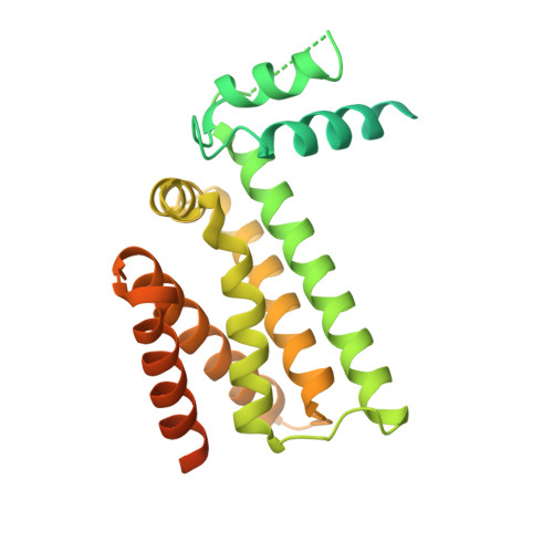
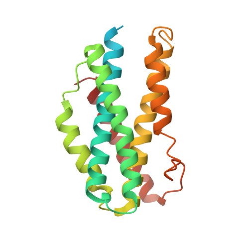
Structure and functionality of a multimeric human COQ7:COQ9 complex.

Manicki, M.Aydin, H.Abriata, L.A.Overmyer, K.A.Guerra, R.M.Coon, J.J.Dal Peraro, M.Frost, A.Pagliarini, D.J.

(2022) Mol Cell 82: 4307-4323.e10

  • DOI: https://doi.org/10.1016/j.molcel.2022.10.003
  • Primary Citation of Related Structures:  
    7SSP, 7SSS

  • PubMed Abstract: 

    Coenzyme Q (CoQ) is a redox-active lipid essential for core metabolic pathways and antioxidant defense. CoQ is synthesized upon the mitochondrial inner membrane by an ill-defined "complex Q" metabolon. Here, we present structure-function analyses of a lipid-, substrate-, and NADH-bound complex comprising two complex Q subunits: the hydroxylase COQ7 and the lipid-binding protein COQ9. We reveal that COQ7 adopts a ferritin-like fold with a hydrophobic channel whose substrate-binding capacity is enhanced by COQ9. Using molecular dynamics, we further show that two COQ7:COQ9 heterodimers form a curved tetramer that deforms the membrane, potentially opening a pathway for the CoQ intermediates to translocate from the bilayer to the proteins' lipid-binding sites. Two such tetramers assemble into a soluble octamer with a pseudo-bilayer of lipids captured within. Together, these observations indicate that COQ7 and COQ9 cooperate to access hydrophobic precursors within the membrane and coordinate subsequent synthesis steps toward producing CoQ.


  • Organizational Affiliation

    Department of Cell Biology and Physiology, Washington University School of Medicine, St. Louis, MO 63110, USA; Morgridge Institute for Research, Madison, WI 53715, USA.


Macromolecules
Find similar proteins by:  (by identity cutoff)  |  3D Structure
Entity ID: 1
MoleculeChains Sequence LengthOrganismDetailsImage
Ubiquinone biosynthesis protein COQ9, mitochondrial
A, B, C, D
318Homo sapiensMutation(s): 0 
Gene Names: COQ9C16orf49HSPC326PSEC0129
UniProt & NIH Common Fund Data Resources
Find proteins for O75208 (Homo sapiens)
Explore O75208 
Go to UniProtKB:  O75208
PHAROS:  O75208
GTEx:  ENSG00000088682 
Entity Groups  
Sequence Clusters30% Identity50% Identity70% Identity90% Identity95% Identity100% Identity
UniProt GroupO75208
Sequence Annotations
Expand
  • Reference Sequence
Find similar proteins by:  (by identity cutoff)  |  3D Structure
Entity ID: 2
MoleculeChains Sequence LengthOrganismDetailsImage
5-demethoxyubiquinone hydroxylase, mitochondrial
E, F, G, H
217Homo sapiensMutation(s): 0 
Gene Names: COQ7
EC: 1.14.99.60
UniProt & NIH Common Fund Data Resources
Find proteins for Q99807 (Homo sapiens)
Explore Q99807 
Go to UniProtKB:  Q99807
PHAROS:  Q99807
GTEx:  ENSG00000167186 
Entity Groups  
Sequence Clusters30% Identity50% Identity70% Identity90% Identity95% Identity100% Identity
UniProt GroupQ99807
Sequence Annotations
Expand
  • Reference Sequence
Small Molecules
Ligands 3 Unique
IDChains Name / Formula / InChI Key2D Diagram3D Interactions
PEV (Subject of Investigation/LOI)
Query on PEV

Download Ideal Coordinates CCD File 
AA [auth E]
BA [auth E]
DA [auth F]
EA [auth F]
FA [auth F]
AA [auth E],
BA [auth E],
DA [auth F],
EA [auth F],
FA [auth F],
GA [auth F],
HA [auth F],
I [auth A],
J [auth A],
JA [auth G],
K [auth A],
L [auth B],
LA [auth G],
M [auth B],
MA [auth G],
NA [auth G],
O [auth C],
OA [auth H],
P [auth C],
PA [auth H],
Q [auth C],
R [auth C],
RA [auth H],
S [auth C],
SA [auth H],
T [auth D],
TA [auth H],
U [auth D],
V [auth D],
W [auth D],
X [auth E],
Y [auth E]
(1S)-2-{[(2-AMINOETHOXY)(HYDROXY)PHOSPHORYL]OXY}-1-[(PALMITOYLOXY)METHYL]ETHYL STEARATE
C39 H78 N O8 P
RPJZYOHZALDGKI-QNGWXLTQSA-N
NAD
Query on NAD

Download Ideal Coordinates CCD File 
CA [auth E],
IA [auth F]
NICOTINAMIDE-ADENINE-DINUCLEOTIDE
C21 H27 N7 O14 P2
BAWFJGJZGIEFAR-NNYOXOHSSA-N
8PP (Subject of Investigation/LOI)
Query on 8PP

Download Ideal Coordinates CCD File 
KA [auth G],
N [auth B],
QA [auth H],
Z [auth E]
2-[(2E,6E,10E,14E,18E,22E,26E)-3,7,11,15,19,23,27,31-OCTAMETHYLDOTRIACONTA-2,6,10,14,18,22,26,30-OCTAENYL]PHENOL
C46 H70 O
VUNQJPPPTJIREN-CMAXTTDKSA-N
Experimental Data & Validation

Experimental Data

  • Method: ELECTRON MICROSCOPY
  • Resolution: 2.40 Å
  • Aggregation State: PARTICLE 
  • Reconstruction Method: SINGLE PARTICLE 

Structure Validation

View Full Validation Report


Entry History & Funding Information

Deposition Data


Funding OrganizationLocationGrant Number
National Institutes of Health/National Institute of General Medical Sciences (NIH/NIGMS)United States5R01GM127673-04

Revision History  (Full details and data files)

  • Version 1.0: 2022-11-02
    Type: Initial release
  • Version 1.1: 2022-11-09
    Changes: Database references
  • Version 1.2: 2022-11-30
    Changes: Database references