


Crystal structure of IS11, a thermophilic esterase
Yakunin, A.F.To be published.
Experimental Data Snapshot
Starting Model: in silico
View more details
wwPDB Validation 3D Report Full Report

Entity ID: 1 | |||||
|---|---|---|---|---|---|
| Molecule | Chains | Sequence Length | Organism | Details | Image |
| IS11 | 456 | Dehalococcoidia bacterium | Mutation(s): 0 | 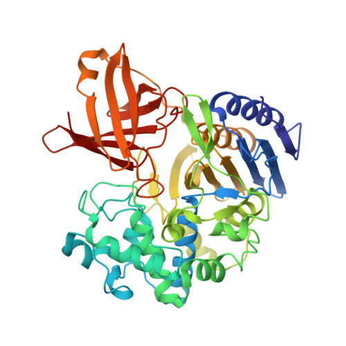 | |
| Length ( Å ) | Angle ( ˚ ) |
|---|---|
| a = 50.216 | α = 90 |
| b = 103.359 | β = 103.481 |
| c = 94.857 | γ = 90 |
| Software Name | Purpose |
|---|---|
| PHENIX | refinement |
| HKL-3000 | data reduction |
| HKL-3000 | data scaling |
| PHENIX | phasing |
| PHENIX | model building |
| Coot | model building |
| Funding Organization | Location | Grant Number |
|---|---|---|
| Not funded | -- |


