7SN6

U2AF65 UHM BOUND TO SF3B155 ULM5


Experimental Data Snapshot

  • Method: X-RAY DIFFRACTION
  • Resolution: 1.80 Å
  • R-Value Free: 0.217 
  • R-Value Work: 0.180 
  • R-Value Observed: 0.183 

wwPDB Validation   3D Report Full Report


This is version 1.2 of the entry. See complete history


Literature

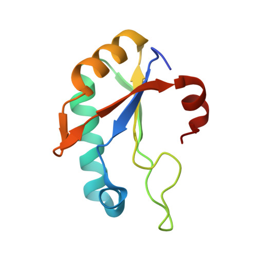
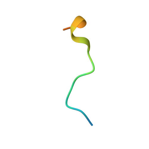
A UHM-ULM interface with unusual structural features contributes to U2AF2 and SF3B1 association for pre-mRNA splicing.

Galardi, J.W.Bela, V.N.Jeffery, N.He, X.Glasser, E.Loerch, S.Jenkins, J.L.Pulvino, M.J.Boutz, P.L.Kielkopf, C.L.

(2022) J Biol Chem 298: 102224-102224

  • DOI: https://doi.org/10.1016/j.jbc.2022.102224
  • Primary Citation of Related Structures:  
    7SN6

  • PubMed Abstract: 

    During spliceosome assembly, the 3' splice site is recognized by sequential U2AF2 complexes, first with Splicing Factor 1 (SF1) and second by the SF3B1 subunit of the U2 small nuclear ribonuclear protein particle. The U2AF2-SF1 interface is well characterized, comprising a U2AF homology motif (UHM) of U2AF2 bound to a U2AF ligand motif (ULM) of SF1. However, the structure of the U2AF2-SF3B1 interface and its importance for pre-mRNA splicing are unknown. To address this knowledge gap, we determined the crystal structure of the U2AF2 UHM bound to a SF3B1 ULM site at 1.8-Å resolution. We discovered a distinctive trajectory of the SF3B1 ULM across the U2AF2 UHM surface, which differs from prior UHM/ULM structures and is expected to modulate the orientations of the full-length proteins. We established that the binding affinity of the U2AF2 UHM for the cocrystallized SF3B1 ULM rivals that of a nearly full-length U2AF2 protein for an N-terminal SF3B1 region. An additional SF3B6 subunit had no detectable effect on the U2AF2-SF3B1 binding affinities. We further showed that key residues at the U2AF2 UHM-SF3B1 ULM interface contribute to coimmunoprecipitation of the splicing factors. Moreover, disrupting the U2AF2-SF3B1 interface changed splicing of representative human transcripts. From analysis of genome-wide data, we found that many of the splice sites coregulated by U2AF2 and SF3B1 differ from those coregulated by U2AF2 and SF1. Taken together, these findings support distinct structural and functional roles for the U2AF2-SF1 and U2AF2-SF3B1 complexes during the pre-mRNA splicing process.


  • Organizational Affiliation

    Center for RNA Biology, Department of Biochemistry and Biophysics, University of Rochester School of Medicine and Dentistry, Rochester, New York, USA.


Macromolecules
Find similar proteins by:  (by identity cutoff)  |  3D Structure
Entity ID: 1
MoleculeChains Sequence LengthOrganismDetailsImage
Splicing factor U2AF 65 kDa subunit
A, B
106Homo sapiensMutation(s): 0 
Gene Names: U2AF2U2AF65
UniProt & NIH Common Fund Data Resources
Find proteins for P26368 (Homo sapiens)
Explore P26368 
Go to UniProtKB:  P26368
PHAROS:  P26368
GTEx:  ENSG00000063244 
Entity Groups  
Sequence Clusters30% Identity50% Identity70% Identity90% Identity95% Identity100% Identity
UniProt GroupP26368
Sequence Annotations
Expand
  • Reference Sequence

Find similar proteins by:  Sequence   |   3D Structure  

Entity ID: 2
MoleculeChains Sequence LengthOrganismDetailsImage
Splicing factor 3B subunit 1
C, D
19Homo sapiensMutation(s): 0 
UniProt & NIH Common Fund Data Resources
Find proteins for O75533 (Homo sapiens)
Explore O75533 
Go to UniProtKB:  O75533
PHAROS:  O75533
GTEx:  ENSG00000115524 
Entity Groups  
Sequence Clusters30% Identity50% Identity70% Identity90% Identity95% Identity100% Identity
UniProt GroupO75533
Sequence Annotations
Expand
  • Reference Sequence
Small Molecules
Ligands 1 Unique
IDChains Name / Formula / InChI Key2D Diagram3D Interactions
IPA
Query on IPA

Download Ideal Coordinates CCD File 
E [auth B]ISOPROPYL ALCOHOL
C3 H8 O
KFZMGEQAYNKOFK-UHFFFAOYSA-N
Experimental Data & Validation

Experimental Data

  • Method: X-RAY DIFFRACTION
  • Resolution: 1.80 Å
  • R-Value Free: 0.217 
  • R-Value Work: 0.180 
  • R-Value Observed: 0.183 
  • Space Group: C 1 2 1
Unit Cell:
Length ( Å )Angle ( ˚ )
a = 76.163α = 90
b = 63.156β = 94.37
c = 49.387γ = 90
Software Package:
Software NamePurpose
PHENIXrefinement
PDB_EXTRACTdata extraction
XDSdata reduction
Aimlessdata scaling
PHASERphasing

Structure Validation

View Full Validation Report


Entry History & Funding Information

Deposition Data


Funding OrganizationLocationGrant Number
National Institutes of Health/National Institute of General Medical Sciences (NIH/NIGMS)United StatesGM070503-15

Revision History  (Full details and data files)

  • Version 1.0: 2022-07-27
    Type: Initial release
  • Version 1.1: 2022-08-24
    Changes: Database references
  • Version 1.2: 2023-10-18
    Changes: Data collection, Refinement description