7SMZ

X-ray crystal structure of CYP142A3 from Mycobacterium Marinum in complex with 4-cholesten-3-one


Experimental Data Snapshot

  • Method: X-RAY DIFFRACTION
  • Resolution: 2.04 Å
  • R-Value Free: 0.225 
  • R-Value Work: 0.180 
  • R-Value Observed: 0.182 

wwPDB Validation   3D Report Full Report


Ligand Structure Quality Assessment 


This is version 1.1 of the entry. See complete history


Literature

The Structures of the Steroid Binding CYP142 Cytochrome P450 Enzymes from Mycobacterium ulcerans and Mycobacterium marinum.

Ghith, A.Doherty, D.Z.Bruning, J.B.Russell, R.A.De Voss, J.J.Bell, S.G.

(2022) ACS Infect Dis 8: 1606-1617

  • DOI: https://doi.org/10.1021/acsinfecdis.2c00215
  • Primary Citation of Related Structures:  
    7SH5, 7SMZ, 7TLO

  • PubMed Abstract: 

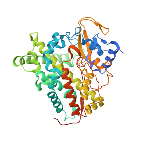
    The steroid binding CYP142 cytochrome P450 enzymes of Mycobacterium species are involved in the metabolism of cholesterol and its derivatives. The equivalent enzyme from Mycobacterium ulcerans was studied to compare the degree of functional conservation between members of this CYP family. We compared substrate binding of the CYP142A3 enzymes of M. ulcerans and M. marinum and CYP142A1 from M. tuberculosis using UV-vis spectroscopy. The catalytic oxidation of cholesterol derivatives by all three enzymes was undertaken. Both CYP142A3 enzymes were structurally characterized by X-ray crystallography. The amino acid sequences of the CYP142A3 enzymes are more similar to CYP142A1 from M. tuberculosis than CYP142A2 from Mycolicibacterium smegmatis . Both CYP142A3 enzymes have substrate binding properties, which are more resemblant to CYP142A1 than CYP142A2. The cholest-4-en-3-one-bound X-ray crystal structure of both CYP142A3 enzymes were determined at a resolution of <1.8 Å, revealing the substrate binding mode at a high level of detail. The structures of the cholest-4-en-3-one binding CYP142 enzymes from M. ulcerans and M. marinum demonstrate how the steroid binds in the active site of these enzymes. They provide an explanation for the high selectivity of the enzyme for terminal methyl C-H bond oxidation to form 26-hydroxy derivatives. These enzymes in pathogenic Mycobacterium species are candidates for inhibition. The work here demonstrates that similar drug molecules could target these CYP142 enzymes from different species in order to combat Buruli ulcer or tuberculosis.


  • Organizational Affiliation

    Department of Chemistry, University of Adelaide, Adelaide, SA 5005, Australia.


Macromolecules
Find similar proteins by:  (by identity cutoff)  |  3D Structure
Entity ID: 1
MoleculeChains Sequence LengthOrganismDetailsImage
Cytochrome P450 142A3
A, B
405Mycobacterium marinum ATCC BAA-535Mutation(s): 0 
Gene Names: cyp142MMRN_53720
UniProt
Find proteins for B2HI07 (Mycobacterium marinum (strain ATCC BAA-535 / M))
Explore B2HI07 
Go to UniProtKB:  B2HI07
Entity Groups  
Sequence Clusters30% Identity50% Identity70% Identity90% Identity95% Identity100% Identity
UniProt GroupB2HI07
Sequence Annotations
Expand
  • Reference Sequence
Small Molecules
Ligands 4 Unique
IDChains Name / Formula / InChI Key2D Diagram3D Interactions
HEM
Query on HEM

Download Ideal Coordinates CCD File 
D [auth A],
J [auth B]
PROTOPORPHYRIN IX CONTAINING FE
C34 H32 Fe N4 O4
KABFMIBPWCXCRK-RGGAHWMASA-L
K2B (Subject of Investigation/LOI)
Query on K2B

Download Ideal Coordinates CCD File 
C [auth A],
K [auth B]
(8ALPHA,9BETA)-CHOLEST-4-EN-3-ONE
C27 H44 O
NYOXRYYXRWJDKP-GYKMGIIDSA-N
ACT
Query on ACT

Download Ideal Coordinates CCD File 
E [auth A]
F [auth A]
G [auth A]
H [auth A]
L [auth B]
E [auth A],
F [auth A],
G [auth A],
H [auth A],
L [auth B],
M [auth B],
N [auth B],
O [auth B]
ACETATE ION
C2 H3 O2
QTBSBXVTEAMEQO-UHFFFAOYSA-M
NA
Query on NA

Download Ideal Coordinates CCD File 
I [auth A],
P [auth B],
Q [auth B],
R [auth B]
SODIUM ION
Na
FKNQFGJONOIPTF-UHFFFAOYSA-N
Experimental Data & Validation

Experimental Data

  • Method: X-RAY DIFFRACTION
  • Resolution: 2.04 Å
  • R-Value Free: 0.225 
  • R-Value Work: 0.180 
  • R-Value Observed: 0.182 
  • Space Group: P 1
Unit Cell:
Length ( Å )Angle ( ˚ )
a = 50.313α = 82.32
b = 50.395β = 75.59
c = 82.577γ = 72.05
Software Package:
Software NamePurpose
PHENIXrefinement
Aimlessdata scaling
PDB_EXTRACTdata extraction
XDSdata reduction
PHASERphasing

Structure Validation

View Full Validation Report


Ligand Structure Quality Assessment 


Entry History & Funding Information

Deposition Data


Funding OrganizationLocationGrant Number
Australian Research Council (ARC)AustraliaDP210103970

Revision History  (Full details and data files)

  • Version 1.0: 2022-09-14
    Type: Initial release
  • Version 1.1: 2023-10-18
    Changes: Data collection, Refinement description