7SL8

CryoEM structure of SGLT1 at 3.4 A resolution


Experimental Data Snapshot

  • Method: ELECTRON MICROSCOPY
  • Resolution: 3.40 Å
  • Aggregation State: PARTICLE 
  • Reconstruction Method: SINGLE PARTICLE 

wwPDB Validation   3D Report Full Report


This is version 1.2 of the entry. See complete history


Literature

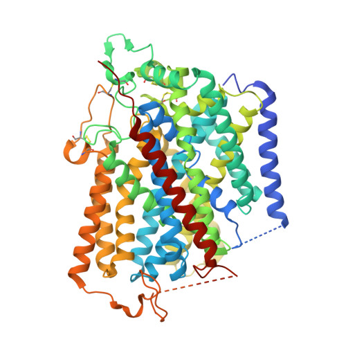
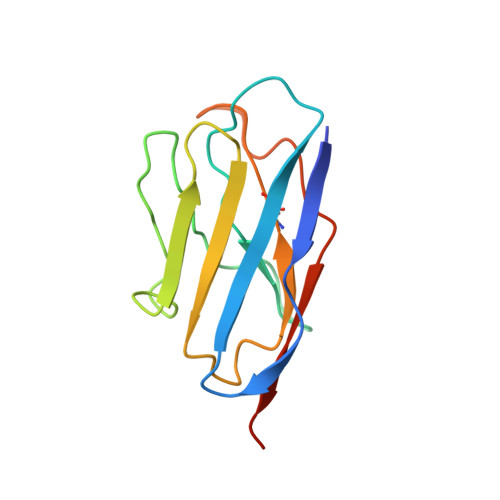
Structure and mechanism of the SGLT family of glucose transporters.

Han, L.Qu, Q.Aydin, D.Panova, O.Robertson, M.J.Xu, Y.Dror, R.O.Skiniotis, G.Feng, L.

(2022) Nature 601: 274-279

  • DOI: https://doi.org/10.1038/s41586-021-04211-w
  • Primary Citation of Related Structures:  
    7SL8, 7SL9, 7SLA

  • PubMed Abstract: 

    Glucose is a primary energy source in living cells. The discovery in 1960s that a sodium gradient powers the active uptake of glucose in the intestine 1 heralded the concept of a secondary active transporter that can catalyse the movement of a substrate against an electrochemical gradient by harnessing energy from another coupled substrate. Subsequently, coupled Na + /glucose transport was found to be mediated by sodium-glucose cotransporters 2,3 (SGLTs). SGLTs are responsible for active glucose and galactose absorption in the intestine and for glucose reabsorption in the kidney 4 , and are targeted by multiple drugs to treat diabetes 5 . Several members within the SGLT family transport key metabolites other than glucose 2 . Here we report cryo-electron microscopy structures of the prototypic human SGLT1 and a related monocarboxylate transporter SMCT1 from the same family. The structures, together with molecular dynamics simulations and functional studies, define the architecture of SGLTs, uncover the mechanism of substrate binding and selectivity, and shed light on water permeability of SGLT1. These results provide insights into the multifaceted functions of SGLTs.


  • Organizational Affiliation

    Department of Molecular and Cellular Physiology, Stanford University School of Medicine, Stanford, CA, USA.


Macromolecules
Find similar proteins by:  (by identity cutoff)  |  3D Structure
Entity ID: 1
MoleculeChains Sequence LengthOrganismDetailsImage
Sodium/glucose cotransporter 1673Homo sapiensMutation(s): 0 
Membrane Entity: Yes 
UniProt & NIH Common Fund Data Resources
Find proteins for P13866 (Homo sapiens)
Explore P13866 
Go to UniProtKB:  P13866
PHAROS:  P13866
GTEx:  ENSG00000100170 
Entity Groups  
Sequence Clusters30% Identity50% Identity70% Identity90% Identity95% Identity100% Identity
UniProt GroupP13866
Sequence Annotations
Expand
  • Reference Sequence
Find similar proteins by:  (by identity cutoff)  |  3D Structure
Entity ID: 2
MoleculeChains Sequence LengthOrganismDetailsImage
nanobody Nb1126synthetic constructMutation(s): 0 
Entity Groups  
Sequence Clusters30% Identity50% Identity70% Identity90% Identity95% Identity100% Identity
Sequence Annotations
Expand
  • Reference Sequence
Small Molecules
Ligands 1 Unique
IDChains Name / Formula / InChI Key2D Diagram3D Interactions
CLR (Subject of Investigation/LOI)
Query on CLR

Download Ideal Coordinates CCD File 
C [auth A]CHOLESTEROL
C27 H46 O
HVYWMOMLDIMFJA-DPAQBDIFSA-N
Experimental Data & Validation

Experimental Data

  • Method: ELECTRON MICROSCOPY
  • Resolution: 3.40 Å
  • Aggregation State: PARTICLE 
  • Reconstruction Method: SINGLE PARTICLE 

Structure Validation

View Full Validation Report


Entry History & Funding Information

Deposition Data


Funding OrganizationLocationGrant Number
Not funded--

Revision History  (Full details and data files)

  • Version 1.0: 2021-12-15
    Type: Initial release
  • Version 1.1: 2021-12-22
    Changes: Database references
  • Version 1.2: 2022-01-26
    Changes: Database references