7SJM | pdb_00007sjm

anti-HtrA1 Fab15H6.v4


Experimental Data Snapshot

  • Method: X-RAY DIFFRACTION
  • Resolution: 1.80 Å
  • R-Value Free: 
    0.202 (Depositor), 0.206 (DCC) 
  • R-Value Work: 
    0.171 (Depositor), 0.173 (DCC) 
  • R-Value Observed: 
    0.173 (Depositor) 

Starting Model: experimental
View more details

wwPDB Validation 3D Report Full Report

Validation slider image for 7SJM

This is version 1.3 of the entry. See complete history

Literature

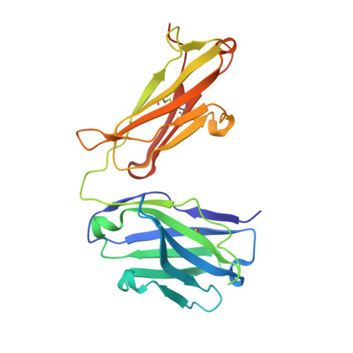
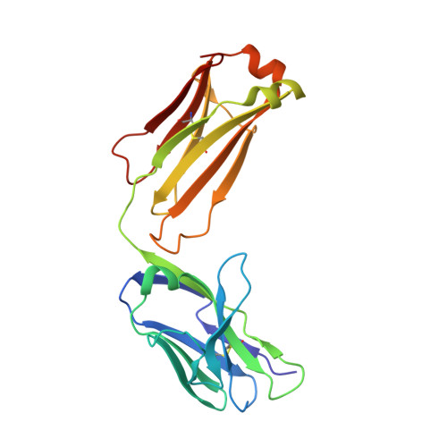
Allosteric inhibition of HTRA1 activity by a conformational lock mechanism to treat age-related macular degeneration.

Gerhardy, S.Ultsch, M.Tang, W.Green, E.Holden, J.K.Li, W.Estevez, A.Arthur, C.Tom, I.Rohou, A.Kirchhofer, D.

(2022) Nat Commun 13: 5222-5222

  • DOI: https://doi.org/10.1038/s41467-022-32760-9
  • Primary Citation Related Structures: 
    7SJM, 7SJN, 7SJO, 7SJP

  • PubMed Abstract: 

    The trimeric serine protease HTRA1 is a genetic risk factor associated with geographic atrophy (GA), a currently untreatable form of age-related macular degeneration. Here, we describe the allosteric inhibition mechanism of HTRA1 by a clinical Fab fragment, currently being evaluated for GA treatment. Using cryo-EM, X-ray crystallography and biochemical assays we identify the exposed LoopA of HTRA1 as the sole Fab epitope, which is approximately 30 Å away from the active site. The cryo-EM structure of the HTRA1:Fab complex in combination with molecular dynamics simulations revealed that Fab binding to LoopA locks HTRA1 in a non-competent conformational state, incapable of supporting catalysis. Moreover, grafting the HTRA1-LoopA epitope onto HTRA2 and HTRA3 transferred the allosteric inhibition mechanism. This suggests a conserved conformational lock mechanism across the HTRA family and a critical role of LoopA for catalysis, which was supported by the reduced activity of HTRA1-3 upon LoopA deletion or perturbation. This study reveals the long-range inhibition mechanism of the clinical Fab and identifies an essential function of the exposed LoopA for activity of HTRA family proteases.


  • Organizational Affiliation
    • Department of Early Discovery Biochemistry, Genentech Inc., San Francisco, CA, USA.

Macromolecule Content 

  • Total Structure Weight: 47.98 kDa 
  • Atom Count: 3,827 
  • Modeled Residue Count: 433 
  • Deposited Residue Count: 440 
  • Unique protein chains: 2

Macromolecules

Find similar proteins by:|  3D Structure
Entity ID: 1
MoleculeChains  Sequence LengthOrganismDetailsImage
Heavy ChainA [auth H]227Homo sapiensMutation(s): 0 
Find similar proteins by:|  3D Structure
Entity ID: 2
MoleculeChains  Sequence LengthOrganismDetailsImage
Light ChainB [auth L]213Homo sapiensMutation(s): 0 

Experimental Data & Validation

Experimental Data

  • Method: X-RAY DIFFRACTION
  • Resolution: 1.80 Å
  • R-Value Free:  0.202 (Depositor), 0.206 (DCC) 
  • R-Value Work:  0.171 (Depositor), 0.173 (DCC) 
  • R-Value Observed: 0.173 (Depositor) 
Space Group: C 1 2 1
Unit Cell:
Length ( Å )Angle ( ˚ )
a = 154.118α = 90
b = 60.338β = 97.32
c = 53.444γ = 90
Software Package:
Software NamePurpose
BUSTERrefinement
PDB_EXTRACTdata extraction
XDSdata reduction
autoPROCdata scaling
PHASERphasing

Structure Validation

View Full Validation Report


Entry History 

& Funding Information

Deposition Data


Funding OrganizationLocationGrant Number
Not funded--

Revision History  (Full details and data files)

  • Version 1.0: 2022-09-07
    Type: Initial release
  • Version 1.1: 2022-09-21
    Changes: Database references
  • Version 1.2: 2023-10-18
    Changes: Data collection, Refinement description
  • Version 1.3: 2024-11-13
    Changes: Structure summary