7SJ8

13pf wildtype microtubule from recombinant human tubulin decorated with kinesin


Experimental Data Snapshot

  • Method: ELECTRON MICROSCOPY
  • Resolution: 3.60 Å
  • Aggregation State: HELICAL ARRAY 
  • Reconstruction Method: HELICAL 

wwPDB Validation   3D Report Full Report


This is version 1.0 of the entry. See complete history


Literature

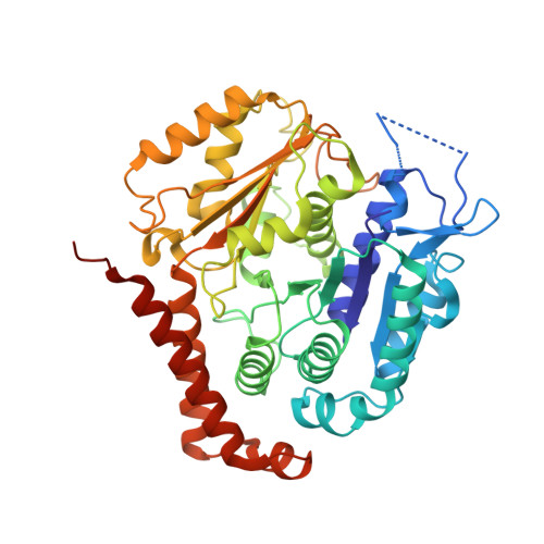
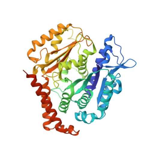
Structural transitions in the GTP cap visualized by cryo-electron microscopy of catalytically inactive microtubules.

LaFrance, B.J.Roostalu, J.Henkin, G.Greber, B.J.Zhang, R.Normanno, D.McCollum, C.O.Surrey, T.Nogales, E.

(2022) Proc Natl Acad Sci U S A 119

  • DOI: https://doi.org/10.1073/pnas.2114994119
  • Primary Citation of Related Structures:  
    7SJ7, 7SJ8, 7SJ9, 7SJA

  • PubMed Abstract: 

    Microtubules (MTs) are polymers of αβ-tubulin heterodimers that stochastically switch between growth and shrinkage phases. This dynamic instability is critically important for MT function. It is believed that GTP hydrolysis within the MT lattice is accompanied by destabilizing conformational changes and that MT stability depends on a transiently existing GTP cap at the growing MT end. Here, we use cryo-electron microscopy and total internal reflection fluorescence microscopy of GTP hydrolysis-deficient MTs assembled from mutant recombinant human tubulin to investigate the structure of a GTP-bound MT lattice. We find that the GTP-MT lattice of two mutants in which the catalytically active glutamate in α-tubulin was substituted by inactive amino acids (E254A and E254N) is remarkably plastic. Undecorated E254A and E254N MTs with 13 protofilaments both have an expanded lattice but display opposite protofilament twists, making these lattices distinct from the compacted lattice of wild-type GDP-MTs. End-binding proteins of the EB family have the ability to compact both mutant GTP lattices and to stabilize a negative twist, suggesting that they promote this transition also in the GTP cap of wild-type MTs, thereby contributing to the maturation of the MT structure. We also find that the MT seam appears to be stabilized in mutant GTP-MTs and destabilized in GDP-MTs, supporting the proposal that the seam plays an important role in MT stability. Together, these structures of catalytically inactive MTs add mechanistic insight into the GTP state of MTs, the stability of the GTP- and GDP-bound lattice, and our overall understanding of MT dynamic instability.


  • Organizational Affiliation

    Department of Molecular and Cell Biology, University of California, Berkeley, CA 94720.


Macromolecules
Find similar proteins by:  (by identity cutoff)  |  3D Structure
Entity ID: 1
MoleculeChains Sequence LengthOrganismDetailsImage
Tubulin alpha-1B chain457Homo sapiensMutation(s): 0 
Gene Names: TUBA1B
UniProt & NIH Common Fund Data Resources
Find proteins for P68363 (Homo sapiens)
Explore P68363 
Go to UniProtKB:  P68363
PHAROS:  P68363
GTEx:  ENSG00000123416 
Entity Groups  
Sequence Clusters30% Identity50% Identity70% Identity90% Identity95% Identity100% Identity
UniProt GroupP68363
Sequence Annotations
Expand
  • Reference Sequence
Find similar proteins by:  (by identity cutoff)  |  3D Structure
Entity ID: 2
MoleculeChains Sequence LengthOrganismDetailsImage
Tubulin beta-3 chain456Homo sapiensMutation(s): 0 
Gene Names: TUBB3TUBB4
UniProt & NIH Common Fund Data Resources
Find proteins for Q13509 (Homo sapiens)
Explore Q13509 
Go to UniProtKB:  Q13509
PHAROS:  Q13509
GTEx:  ENSG00000258947 
Entity Groups  
Sequence Clusters30% Identity50% Identity70% Identity90% Identity95% Identity100% Identity
UniProt GroupQ13509
Sequence Annotations
Expand
  • Reference Sequence
Small Molecules
Ligands 3 Unique
IDChains Name / Formula / InChI Key2D Diagram3D Interactions
GTP (Subject of Investigation/LOI)
Query on GTP

Download Ideal Coordinates CCD File 
M [auth A]
O [auth C]
Q [auth E]
S [auth J]
U [auth K]
M [auth A],
O [auth C],
Q [auth E],
S [auth J],
U [auth K],
W [auth L]
GUANOSINE-5'-TRIPHOSPHATE
C10 H16 N5 O14 P3
XKMLYUALXHKNFT-UUOKFMHZSA-N
GDP (Subject of Investigation/LOI)
Query on GDP

Download Ideal Coordinates CCD File 
AA [auth G]
BA [auth H]
CA [auth I]
DA [auth B]
Y [auth F]
AA [auth G],
BA [auth H],
CA [auth I],
DA [auth B],
Y [auth F],
Z [auth D]
GUANOSINE-5'-DIPHOSPHATE
C10 H15 N5 O11 P2
QGWNDRXFNXRZMB-UUOKFMHZSA-N
MG (Subject of Investigation/LOI)
Query on MG

Download Ideal Coordinates CCD File 
N [auth A]
P [auth C]
R [auth E]
T [auth J]
V [auth K]
N [auth A],
P [auth C],
R [auth E],
T [auth J],
V [auth K],
X [auth L]
MAGNESIUM ION
Mg
JLVVSXFLKOJNIY-UHFFFAOYSA-N
Experimental Data & Validation

Experimental Data

  • Method: ELECTRON MICROSCOPY
  • Resolution: 3.60 Å
  • Aggregation State: HELICAL ARRAY 
  • Reconstruction Method: HELICAL 
EM Software:
TaskSoftware PackageVersion
MODEL REFINEMENTPHENIXdev-2883
RECONSTRUCTIONFREALIGN9.11

Structure Validation

View Full Validation Report


Entry History & Funding Information

Deposition Data


Funding OrganizationLocationGrant Number
Howard Hughes Medical Institute (HHMI)United States--

Revision History  (Full details and data files)

  • Version 1.0: 2022-01-19
    Type: Initial release