7SIP

Structure of shaker-IR


Experimental Data Snapshot

  • Method: ELECTRON MICROSCOPY
  • Resolution: 3.00 Å
  • Aggregation State: PARTICLE 
  • Reconstruction Method: SINGLE PARTICLE 

wwPDB Validation   3D Report Full Report


This is version 1.0 of the entry. See complete history


Literature

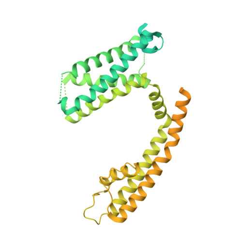
Structure of the Shaker Kv channel and mechanism of slow C-type inactivation.

Tan, X.F.Bae, C.Stix, R.Fernandez-Marino, A.I.Huffer, K.Chang, T.H.Jiang, J.Faraldo-Gomez, J.D.Swartz, K.J.

(2022) Sci Adv 8: eabm7814-eabm7814

  • DOI: https://doi.org/10.1126/sciadv.abm7814
  • Primary Citation of Related Structures:  
    7SIP, 7SJ1

  • PubMed Abstract: 

    Voltage-activated potassium (Kv) channels open upon membrane depolarization and proceed to spontaneously inactivate. Inactivation controls neuronal firing rates and serves as a form of short-term memory and is implicated in various human neurological disorders. Here, we use high-resolution cryo-electron microscopy and computer simulations to determine one of the molecular mechanisms underlying this physiologically crucial process. Structures of the activated Shaker Kv channel and of its W434F mutant in lipid bilayers demonstrate that C-type inactivation entails the dilation of the ion selectivity filter and the repositioning of neighboring residues known to be functionally critical. Microsecond-scale molecular dynamics trajectories confirm that these changes inhibit rapid ion permeation through the channel. This long-sought breakthrough establishes how eukaryotic K + channels self-regulate their functional state through the plasticity of their selectivity filters.


  • Organizational Affiliation

    Molecular Physiology and Biophysics Section, Porter Neuroscience Research Center, National Institute of Neurological Disorders and Stroke, National Institutes of Health, Bethesda, MD 20892, USA.


Macromolecules
Find similar proteins by:  (by identity cutoff)  |  3D Structure
Entity ID: 1
MoleculeChains Sequence LengthOrganismDetailsImage
Potassium voltage-gated channel protein Shaker
A, B, C, D
617Drosophila melanogasterMutation(s): 0 
Gene Names: ShmnsCG12348
Membrane Entity: Yes 
UniProt
Find proteins for P08510 (Drosophila melanogaster)
Explore P08510 
Go to UniProtKB:  P08510
Entity Groups  
Sequence Clusters30% Identity50% Identity70% Identity90% Identity95% Identity100% Identity
UniProt GroupP08510
Sequence Annotations
Expand
  • Reference Sequence
Small Molecules
Ligands 2 Unique
IDChains Name / Formula / InChI Key2D Diagram3D Interactions
POV
Query on POV

Download Ideal Coordinates CCD File 
AA [auth C]
BA [auth C]
CA [auth C]
DA [auth C]
E [auth A]
AA [auth C],
BA [auth C],
CA [auth C],
DA [auth C],
E [auth A],
EA [auth C],
F [auth A],
FA [auth C],
G [auth A],
GA [auth C],
H [auth A],
HA [auth D],
I [auth A],
IA [auth D],
J [auth A],
JA [auth D],
K [auth A],
KA [auth D],
L [auth A],
LA [auth D],
M [auth A],
MA [auth D],
NA [auth D],
R [auth B],
S [auth B],
T [auth B],
U [auth B],
V [auth B],
W [auth B],
X [auth B],
Y [auth B],
Z [auth C]
(2S)-3-(hexadecanoyloxy)-2-[(9Z)-octadec-9-enoyloxy]propyl 2-(trimethylammonio)ethyl phosphate
C42 H82 N O8 P
WTJKGGKOPKCXLL-PFDVCBLKSA-N
K (Subject of Investigation/LOI)
Query on K

Download Ideal Coordinates CCD File 
N [auth A],
O [auth A],
P [auth A],
Q [auth A]
POTASSIUM ION
K
NPYPAHLBTDXSSS-UHFFFAOYSA-N
Experimental Data & Validation

Experimental Data

  • Method: ELECTRON MICROSCOPY
  • Resolution: 3.00 Å
  • Aggregation State: PARTICLE 
  • Reconstruction Method: SINGLE PARTICLE 

Structure Validation

View Full Validation Report


Entry History & Funding Information

Deposition Data


Funding OrganizationLocationGrant Number
National Institutes of Health/National Institute of Neurological Disorders and Stroke (NIH/NINDS)United States--

Revision History  (Full details and data files)

  • Version 1.0: 2022-03-30
    Type: Initial release