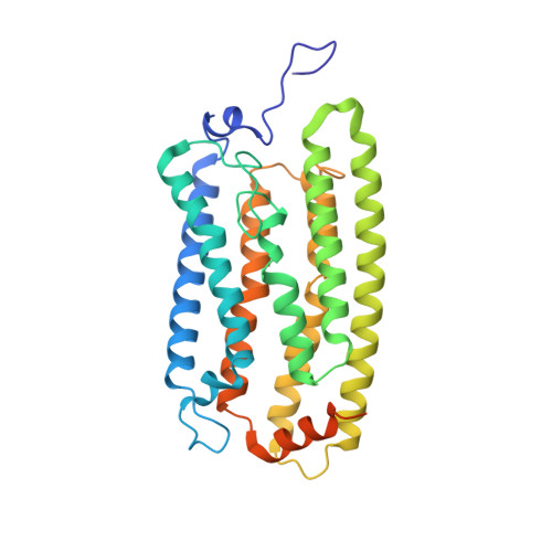
Cryo-EM structures of the channelrhodopsin ChRmine in lipid nanodiscs.
Tucker, K., Sridharan, S., Adesnik, H., Brohawn, S.G.(2022) Nat Commun 13: 4842-4842
- PubMed: 35977941 Search on PubMedSearch on PubMed Central
- DOI: https://doi.org/10.1038/s41467-022-32441-7
- Primary Citation Related Structures:
7SFJ, 7SFK, 7SHS - PubMed Abstract:
Microbial channelrhodopsins are light-gated ion channels widely used for optogenetic manipulation of neuronal activity. ChRmine is a bacteriorhodopsin-like cation channelrhodopsin (BCCR) more closely related to ion pump rhodopsins than other channelrhodopsins. ChRmine displays unique properties favorable for optogenetics including high light sensitivity, a broad, red-shifted activation spectrum, cation selectivity, and large photocurrents, while its slow closing kinetics impedes some applications. The structural basis for ChRmine function, or that of any other BCCR, is unknown. Here, we present cryo-EM structures of ChRmine in lipid nanodiscs in apo (opsin) and retinal-bound (rhodopsin) forms. The structures reveal an unprecedented trimeric architecture with a lipid filled central pore. Large electronegative cavities on either side of the membrane facilitate high conductance and selectivity for cations over protons. The retinal binding pocket structure suggests channel properties could be tuned with mutations and we identify ChRmine variants with ten-fold decreased and two-fold increased closing rates. A T119A mutant shows favorable properties relative to wild-type and previously reported ChRmine variants for optogenetics. These results provide insight into structural features that generate an ultra-potent microbial opsin and provide a platform for rational engineering of channelrhodopsins with improved properties that could expand the scale, depth, and precision of optogenetic experiments.
- Department of Molecular & Cell Biology, University of California Berkeley, Berkeley, CA, 94720, USA.
Organizational Affiliation: