7SHH | pdb_00007shh

Bacterial cereblon homologue in complex with (R)-3-(4-methoxyphenyl)piperidine-2,6-dione


Experimental Data Snapshot

  • Method: X-RAY DIFFRACTION
  • Resolution: 1.90 Å
  • R-Value Free: 
    0.216 (Depositor), 0.218 (DCC) 
  • R-Value Work: 
    0.192 (Depositor), 0.194 (DCC) 
  • R-Value Observed: 
    0.193 (Depositor) 

wwPDB Validation 3D Report Full Report

Validation slider image for 7SHH

Ligand Structure Quality Assessment 


This is version 1.1 of the entry. See complete history

Literature

Development of Potent and Selective Janus Kinase 2/3 Directing PG-PROTACs.

Alcock, L.J.Chang, Y.Jarusiewicz, J.A.Actis, M.Nithianantham, S.Mayasundari, A.Min, J.Maxwell, D.Hunt, J.Smart, B.Yang, J.J.Nishiguchi, G.Fischer, M.Mullighan, C.G.Rankovic, Z.

(2022) ACS Med Chem Lett 13: 475-482

  • DOI: https://doi.org/10.1021/acsmedchemlett.1c00650
  • Primary Citation Related Structures: 
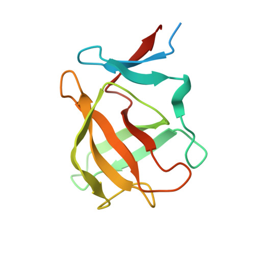
    7SHH

  • PubMed Abstract: 

    Aberrant activation of the JAK-STAT signaling pathway has been implicated in the pathogenesis of a range of hematological malignancies and autoimmune disorders. Here we describe the design, synthesis, and characterization of JAK2/3 PROTACs utilizing a phenyl glutarimide (PG) ligand as the cereblon (CRBN) recruiter. SJ10542 displayed high selectivity over GSPT1 and other members of the JAK family and potency in patient-derived ALL cells containing both JAK2 fusions and CRLF2 rearrangements.


  • Organizational Affiliation
    • Department of Pathology, St. Jude Children's Research Hospital, Memphis, Tennessee 38105, United States.

Macromolecule Content 

  • Total Structure Weight: 41.92 kDa 
  • Atom Count: 2,557 
  • Modeled Residue Count: 311 
  • Deposited Residue Count: 375 
  • Unique protein chains: 1

Macromolecules

Find similar proteins by:|  3D Structure
Entity ID: 1
MoleculeChains  Sequence LengthOrganismDetailsImage
Cereblon isoform 4
A, B, C
125Magnetospirillum gryphiswaldenseMutation(s): 0 
Gene Names: MGR_0879
UniProt
Find proteins for A4TVL0 (Magnetospirillum gryphiswaldense)
Explore A4TVL0 
Go to UniProtKB:  A4TVL0
Entity Groups
Sequence Clusters30% Identity50% Identity70% Identity90% Identity95% Identity100% Identity
UniProt GroupA4TVL0
Sequence Annotations
Expand
Reference Sequence

Experimental Data & Validation

Experimental Data

  • Method: X-RAY DIFFRACTION
  • Resolution: 1.90 Å
  • R-Value Free:  0.216 (Depositor), 0.218 (DCC) 
  • R-Value Work:  0.192 (Depositor), 0.194 (DCC) 
  • R-Value Observed: 0.193 (Depositor) 
Space Group: P 21 21 21
Unit Cell:
Length ( Å )Angle ( ˚ )
a = 56.311α = 90
b = 59.586β = 90
c = 87.706γ = 90
Software Package:
Software NamePurpose
XDSdata reduction
SCALAdata scaling
PHENIXrefinement
PDB_EXTRACTdata extraction
PHENIXphasing

Structure Validation

View Full Validation Report


Ligand Structure Quality Assessment 


Entry History 

& Funding Information

Deposition Data


Funding OrganizationLocationGrant Number
Other private--

Revision History  (Full details and data files)

  • Version 1.0: 2022-04-13
    Type: Initial release
  • Version 1.1: 2024-05-22
    Changes: Data collection, Refinement description