


Structural Basis for Mis18 Complex Assembly: Implications for Centromere Maintenance
Park, S.H., Shimanaka, K., Cho, U.To be published.
Experimental Data Snapshot
Starting Model: experimental
View more details
Entity ID: 1 | |||||
|---|---|---|---|---|---|
| Molecule | Chains | Sequence Length | Organism | Details | Image |
| Protein Mis18-alpha | 114 | Homo sapiens | Mutation(s): 0 Gene Names: MIS18A, C21orf45, C21orf46, FASP1 | 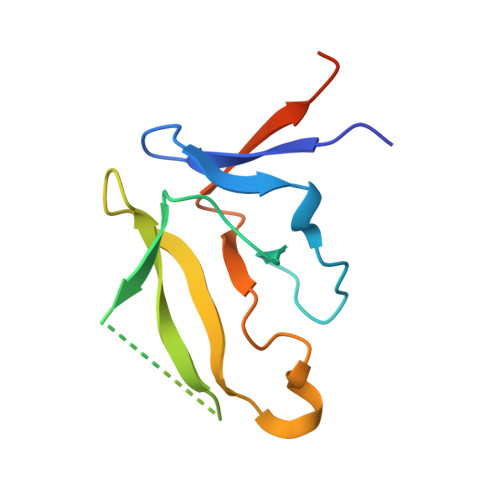 | |
UniProt & NIH Common Fund Data Resources | |||||
PHAROS: Q9NYP9 GTEx: ENSG00000159055 | |||||
Entity Groups | |||||
| Sequence Clusters | 30% Identity50% Identity70% Identity90% Identity95% Identity100% Identity | ||||
| UniProt Group | Q9NYP9 | ||||
Sequence AnnotationsExpand | |||||
Reference Sequence | |||||
| Ligands 2 Unique | |||||
|---|---|---|---|---|---|
| ID | Chains | Name / Formula / InChI Key | 2D Diagram | 3D Interactions | |
| SO4 (Subject of Investigation/LOI) Download:Ideal Coordinates CCD File | K [auth A] L [auth A] O [auth D] P [auth D] R [auth E] | SULFATE ION O4 S QAOWNCQODCNURD-UHFFFAOYSA-L |  | ||
| ZN (Subject of Investigation/LOI) Download:Ideal Coordinates CCD File | I [auth B] J [auth A] M [auth C] N [auth D] Q [auth E] | ZINC ION Zn PTFCDOFLOPIGGS-UHFFFAOYSA-N |  | ||
| Length ( Å ) | Angle ( ˚ ) |
|---|---|
| a = 110.725 | α = 90 |
| b = 114.864 | β = 90 |
| c = 116.27 | γ = 90 |
| Software Name | Purpose |
|---|---|
| PHENIX | refinement |
| HKL-2000 | data reduction |
| HKL-2000 | data scaling |
| PHASER | phasing |
| Funding Organization | Location | Grant Number |
|---|---|---|
| National Institutes of Health/National Institute of Diabetes and Digestive and Kidney Disease (NIH/NIDDK) | United States | DK111465 |


