7SFA

Branchiostoma floridae fluorescent protein LanFP10A2


Experimental Data Snapshot

  • Method: X-RAY DIFFRACTION
  • Resolution: 1.65 Å
  • R-Value Free: 0.197 
  • R-Value Work: 0.165 
  • R-Value Observed: 0.165 

wwPDB Validation   3D Report Full Report


This is version 2.0 of the entry. See complete history


Literature

A novel violet fluorescent protein contains a unique oxidized tyrosine as the simplest chromophore ever reported in fluorescent proteins.

Roldan-Salgado, A.Muslinkina, L.Pletnev, S.Pletneva, N.Pletnev, V.Gaytan, P.

(2022) Protein Sci 31: 688-700

  • DOI: https://doi.org/10.1002/pro.4265
  • Primary Citation of Related Structures:  
    7SF9, 7SFA

  • PubMed Abstract: 

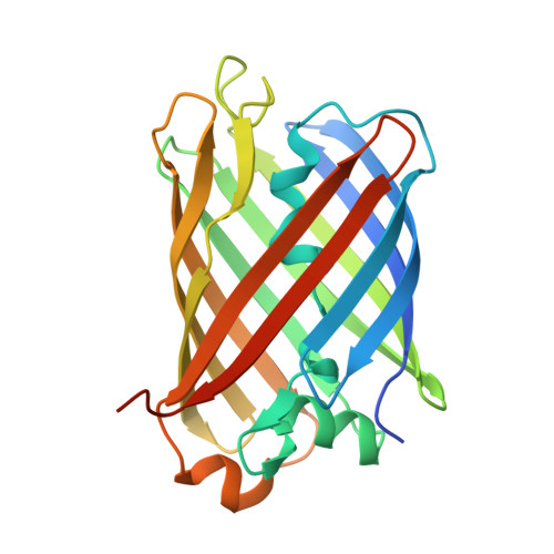
    We describe an engineered violet fluorescent protein from the lancelet Branchiostoma floridae (bfVFP). This is the first example of a GFP-like fluorescent protein with a stable fluorescent chromophore lacking an imidazolinone ring; instead, it consists of oxidized tyrosine 68 flanked by glycine 67 and alanine 69. bfVFP contains the simplest chromophore reported in fluorescent proteins and was generated from the yellow protein lanFP10A2 by two synergetic mutations, S148H and C166I. The chromophore structure was confirmed crystallographically and by high-resolution mass spectrometry. The photophysical characteristics of bfVFP (323/430 nm, quantum yield 0.33, and E c 14,300 M -1  cm -1 ) make it potentially useful for multicolor experiments to expand the excitation range of available FP biomarkers and Förster resonance energy transfer with blue and cyan fluorescent protein acceptors.


  • Organizational Affiliation

    Instituto de Biotecnología, Universidad Nacional Autónoma de México, Cuernavaca, Mexico.


Macromolecules
Find similar proteins by:  (by identity cutoff)  |  3D Structure
Entity ID: 1
MoleculeChains Sequence LengthOrganismDetailsImage
Fluorescent protein lanFP10A2
A, B, C, D
236Branchiostoma floridaeMutation(s): 0 
Gene Names: BRAFLDRAFT_75522
UniProt
Find proteins for C3YRA2 (Branchiostoma floridae)
Explore C3YRA2 
Go to UniProtKB:  C3YRA2
Entity Groups  
Sequence Clusters30% Identity50% Identity70% Identity90% Identity95% Identity100% Identity
UniProt GroupC3YRA2
Sequence Annotations
Expand
  • Reference Sequence
Experimental Data & Validation

Experimental Data

  • Method: X-RAY DIFFRACTION
  • Resolution: 1.65 Å
  • R-Value Free: 0.197 
  • R-Value Work: 0.165 
  • R-Value Observed: 0.165 
  • Space Group: P 2 21 21
Unit Cell:
Length ( Å )Angle ( ˚ )
a = 59.238α = 90
b = 96.217β = 90
c = 158.948γ = 90
Software Package:
Software NamePurpose
HKL-2000data scaling
REFMACrefinement
PDB_EXTRACTdata extraction
HKL-2000data reduction
MOLREPphasing

Structure Validation

View Full Validation Report


Entry History & Funding Information

Deposition Data


Funding OrganizationLocationGrant Number
National Institutes of Health/National Institute Of Allergy and Infectious Diseases (NIH/NIAID)United States--

Revision History  (Full details and data files)

  • Version 1.0: 2022-03-30
    Type: Initial release
  • Version 1.1: 2023-10-18
    Changes: Data collection, Refinement description
  • Version 2.0: 2023-11-15
    Changes: Atomic model, Data collection, Derived calculations