Stabilization of glucose-6-phosphate dehydrogenase oligomers enhances catalytic activity and stability of clinical variants.
Garcia, A.A., Mathews, I.I., Horikoshi, N., Matsui, T., Kaur, M., Wakatsuki, S., Mochly-Rosen, D.(2022) J Biol Chem 298: 101610-101610
- PubMed: 35065072 Search on PubMedSearch on PubMed Central
- DOI: https://doi.org/10.1016/j.jbc.2022.101610
- Primary Citation Related Structures:
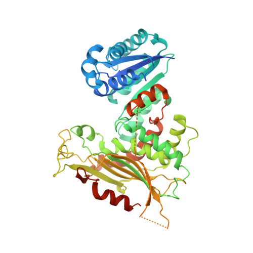
7SEH, 7SEI - PubMed Abstract:
Glucose-6-phosphate dehydrogenase (G6PD) deficiency is a genetic trait that can cause hemolytic anemia. To date, over 150 nonsynonymous mutations have been identified in G6PD, with pathogenic mutations clustering near the dimer and/or tetramer interface and the allosteric NADP + -binding site. Recently, our lab identified a small molecule that activates G6PD variants by stabilizing the allosteric NADP + and dimer complex, suggesting therapeutics that target these regions may improve structural defects. Here, we elucidated the connection between allosteric NADP + binding, oligomerization, and pathogenicity to determine whether oligomer stabilization can be used as a therapeutic strategy for G6PD deficiency (G6PD def ). We first solved the crystal structure for G6PD K403Q , a mutant that mimics the physiological acetylation of wild-type G6PD in erythrocytes and demonstrated that loss of allosteric NADP + binding induces conformational changes in the dimer. These structural changes prevent tetramerization, are unique to Class I variants (the most severe form of G6PD def ), and cause the deactivation and destabilization of G6PD. We also introduced nonnative cysteines at the oligomer interfaces and found that the tetramer complex is more catalytically active and stable than the dimer. Furthermore, stabilizing the dimer and tetramer improved protein stability in clinical variants, regardless of clinical classification, with tetramerization also improving the activity of G6PD K403Q and Class I variants. These findings were validated using enzyme activity and thermostability assays, analytical size-exclusion chromatography (SEC), and SEC coupled with small-angle X-ray scattering (SEC-SAXS). Taken together, our findings suggest a potential therapeutic strategy for G6PD def and provide a foundation for future drug discovery efforts.
- Department of Chemical and Systems Biology, School of Medicine, Stanford University, Stanford, California, USA.
Organizational Affiliation: