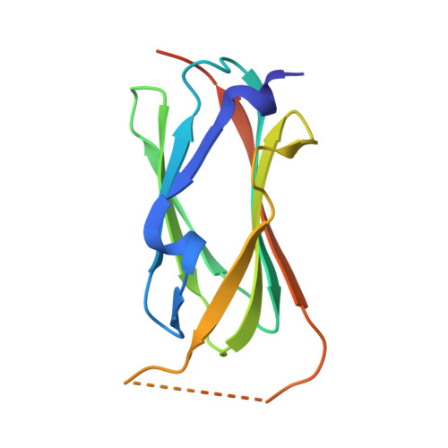
7SC4

filamin complex-1

  • Classification: CELL ADHESION
  • Organism(s): Homo sapiens
  • Expression System: Escherichia phage EcSzw-2
  • Mutation(s): No 

  • Deposited: 2021-09-27 Released: 2023-04-12 
  • Deposition Author(s): Liu, J., Qin, J.
  • Funding Organization(s): National Institutes of Health/National Heart, Lung, and Blood Institute (NIH/NHLBI), American Heart Association

Experimental Data Snapshot

  • Method: X-RAY DIFFRACTION
  • Resolution: 1.85 Å
  • R-Value Free: 0.261 
  • R-Value Work: 0.199 
  • R-Value Observed: 0.202 

wwPDB Validation   3D Report Full Report


This is version 1.1 of the entry. See complete history


Literature

A mechanism of platelet integrin alpha IIb beta 3 outside-in signaling through a novel integrin alpha IIb subunit-filamin-actin linkage.

Liu, J.Lu, F.Ithychanda, S.S.Apostol, M.Das, M.Deshpande, G.Plow, E.F.Qin, J.

(2023) Blood 141: 2629-2641

  • DOI: https://doi.org/10.1182/blood.2022018333
  • Primary Citation of Related Structures:  
    7SC4, 7SFT

  • PubMed Abstract: 

    The communication of talin-activated integrin αIIbβ3 with the cytoskeleton (integrin outside-in signaling) is essential for platelet aggregation, wound healing, and hemostasis. Filamin, a large actin crosslinker and integrin binding partner critical for cell spreading and migration, is implicated as a key regulator of integrin outside-in signaling. However, the current dogma is that filamin, which stabilizes inactive αIIbβ3, is displaced from αIIbβ3 by talin to promote the integrin activation (inside-out signaling), and how filamin further functions remains unresolved. Here, we show that while associating with the inactive αIIbβ3, filamin also associates with the talin-bound active αIIbβ3 to mediate platelet spreading. Fluorescence resonance energy transfer-based analysis reveals that while associating with both αIIb and β3 cytoplasmic tails (CTs) to maintain the inactive αIIbβ3, filamin is spatiotemporally rearranged to associate with αIIb CT alone on activated αIIbβ3. Consistently, confocal cell imaging indicates that integrin α CT-linked filamin gradually delocalizes from the β CT-linked focal adhesion marker-vinculin likely because of the separation of integrin α/β CTs occurring during integrin activation. High-resolution crystal and nuclear magnetic resonance structure determinations unravel that the activated integrin αIIb CT binds to filamin via a striking α-helix→β-strand transition with a strengthened affinity that is dependent on the integrin-activating membrane environment containing enriched phosphatidylinositol 4,5-bisphosphate. These data suggest a novel integrin αIIb CT-filamin-actin linkage that promotes integrin outside-in signaling. Consistently, disruption of such linkage impairs the activation state of αIIbβ3, phosphorylation of focal adhesion kinase/proto-oncogene tyrosine kinase Src, and cell migration. Together, our findings advance the fundamental understanding of integrin outside-in signaling with broad implications in blood physiology and pathology.


  • Organizational Affiliation

    Department of Cardiovascular and Metabolic Sciences, Lerner Research Institute, Cleveland Clinic, Cleveland, OH.


Macromolecules
Find similar proteins by:  (by identity cutoff)  |  3D Structure
Entity ID: 1
MoleculeChains Sequence LengthOrganismDetailsImage
Filamin-A/Integrin alpha-IIb light chain, form 2 chimera
A, B
130Homo sapiensMutation(s): 0 
Gene Names: FLNAFLNFLN1ITGA2BGP2BITGAB
UniProt & NIH Common Fund Data Resources
Find proteins for P08514 (Homo sapiens)
Explore P08514 
Go to UniProtKB:  P08514
PHAROS:  P08514
GTEx:  ENSG00000005961 
Find proteins for P21333 (Homo sapiens)
Explore P21333 
Go to UniProtKB:  P21333
PHAROS:  P21333
GTEx:  ENSG00000196924 
Entity Groups  
Sequence Clusters30% Identity50% Identity70% Identity90% Identity95% Identity100% Identity
UniProt GroupsP21333P08514
Sequence Annotations
Expand
  • Reference Sequence
Small Molecules
Ligands 1 Unique
IDChains Name / Formula / InChI Key2D Diagram3D Interactions
SO4
Query on SO4

Download Ideal Coordinates CCD File 
C [auth B]SULFATE ION
O4 S
QAOWNCQODCNURD-UHFFFAOYSA-L
Experimental Data & Validation

Experimental Data

  • Method: X-RAY DIFFRACTION
  • Resolution: 1.85 Å
  • R-Value Free: 0.261 
  • R-Value Work: 0.199 
  • R-Value Observed: 0.202 
  • Space Group: P 21 2 21
Unit Cell:
Length ( Å )Angle ( ˚ )
a = 50.248α = 90
b = 66.01β = 90
c = 71.889γ = 90
Software Package:
Software NamePurpose
REFMACrefinement
PDB_EXTRACTdata extraction
HKL-3000data reduction
HKL-3000data scaling
PHASERphasing

Structure Validation

View Full Validation Report


Entry History & Funding Information

Deposition Data


Funding OrganizationLocationGrant Number
National Institutes of Health/National Heart, Lung, and Blood Institute (NIH/NHLBI)United StatesR01 HL58758
National Institutes of Health/National Heart, Lung, and Blood Institute (NIH/NHLBI)United StatesP01 HL73311
American Heart AssociationUnited States18CDA34110364

Revision History  (Full details and data files)

  • Version 1.0: 2023-04-12
    Type: Initial release
  • Version 1.1: 2023-10-25
    Changes: Data collection, Database references, Refinement description