7SB7

Crystal structure of T. brucei hypoxanthine guanine phosphoribosyltransferase in complex with (4S,7S)-7-hydroxy-4-((guanin-9-yl)methyl)-2,5-dioxaheptan-1,7-diphosphonate


Experimental Data Snapshot

  • Method: X-RAY DIFFRACTION
  • Resolution: 2.65 Å
  • R-Value Free: 0.253 
  • R-Value Work: 0.196 
  • R-Value Observed: 0.202 

wwPDB Validation   3D Report Full Report


Ligand Structure Quality Assessment 


This is version 1.2 of the entry. See complete history


Literature

Stereo-Defined Acyclic Nucleoside Phosphonates are Selective and Potent Inhibitors of Parasite 6-Oxopurine Phosphoribosyltransferases.

Klejch, T.Keough, D.T.King, G.Dolezelova, E.Cesnek, M.Budesinsky, M.Zikova, A.Janeba, Z.Guddat, L.W.Hockova, D.

(2022) J Med Chem 65: 4030-4057

  • DOI: https://doi.org/10.1021/acs.jmedchem.1c01881
  • Primary Citation of Related Structures:  
    7SAN, 7SB7, 7SCR

  • PubMed Abstract: 

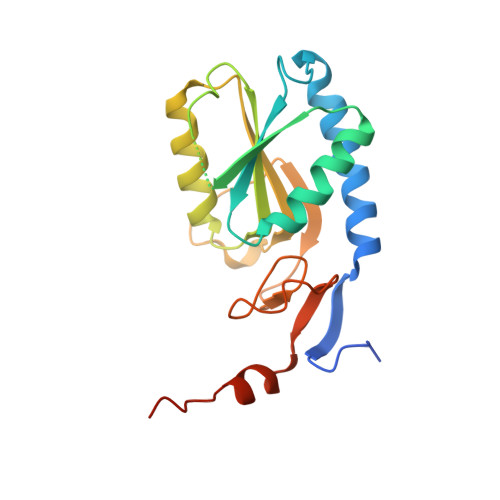
    Pathogens such as Plasmodium and Trypanosoma spp. are unable to synthesize purine nucleobases. They rely on the salvage of these purines and their nucleosides from the host cell to synthesize the purine nucleotides required for DNA/RNA production. The key enzymes in this pathway are purine phosphoribosyltransferases (PRTs). Here, we synthesized 16 novel acyclic nucleoside phosphonates, 12 with a chiral center at C-2', and eight bearing a second chiral center at C-6'. Of these, bisphosphonate ( S , S )- 48 is the most potent inhibitor of the Plasmodium falciparum and P. vivax 6-oxopurine PRTs and the most potent inhibitor of two Trypanosoma brucei ( Tbr ) 6-oxopurine PRTs yet discovered, with K i values as low as 2 nM. Crystal structures of ( S , S )- 48 in complex with human and Tbr 6-oxopurine PRTs show that the inhibitor binds to the enzymes in different conformations, providing an explanation for its potency and selectivity ( i.e. , 35-fold in favor of the parasite enzymes).


  • Organizational Affiliation

    The Institute of Organic Chemistry and Biochemistry of the Czech Academy of Sciences, Prague 6 CZ-16000, Czech Republic.


Macromolecules
Find similar proteins by:  (by identity cutoff)  |  3D Structure
Entity ID: 1
MoleculeChains Sequence LengthOrganismDetailsImage
Hypoxanthine-guanine phosphoribosyltransferase
A, B
216Trypanosoma brucei bruceiMutation(s): 0 
Gene Names: HGPRT
EC: 2.4.2.8
UniProt
Find proteins for Q07010 (Trypanosoma brucei brucei)
Explore Q07010 
Go to UniProtKB:  Q07010
Entity Groups  
Sequence Clusters30% Identity50% Identity70% Identity90% Identity95% Identity100% Identity
UniProt GroupQ07010
Sequence Annotations
Expand
  • Reference Sequence
Small Molecules
Ligands 1 Unique
IDChains Name / Formula / InChI Key2D Diagram3D Interactions
8QI (Subject of Investigation/LOI)
Query on 8QI

Download Ideal Coordinates CCD File 
C [auth A],
D [auth B]
({(2S)-3-(2-amino-6-oxo-1,6-dihydro-9H-purin-9-yl)-2-[(2S)-2-hydroxy-2-phosphonoethoxy]propoxy}methyl)phosphonic acid
C11 H19 N5 O10 P2
BCERVIXRVPEQOK-BQBZGAKWSA-N
Experimental Data & Validation

Experimental Data

  • Method: X-RAY DIFFRACTION
  • Resolution: 2.65 Å
  • R-Value Free: 0.253 
  • R-Value Work: 0.196 
  • R-Value Observed: 0.202 
  • Space Group: C 1 2 1
Unit Cell:
Length ( Å )Angle ( ˚ )
a = 115.908α = 90
b = 93.983β = 109.125
c = 44.274γ = 90
Software Package:
Software NamePurpose
PHENIXrefinement
PHENIXrefinement
XDSdata reduction
Aimlessdata scaling
PHASERphasing

Structure Validation

View Full Validation Report


Ligand Structure Quality Assessment 


Entry History & Funding Information

Deposition Data


Funding OrganizationLocationGrant Number
National Health and Medical Research Council (NHMRC, Australia)Australia1147368

Revision History  (Full details and data files)

  • Version 1.0: 2022-03-02
    Type: Initial release
  • Version 1.1: 2022-03-23
    Changes: Database references
  • Version 1.2: 2023-10-18
    Changes: Data collection, Refinement description