7S9W | pdb_00007s9w

Structure of DrmAB:ADP:DNA complex


Experimental Data Snapshot

  • Method: ELECTRON MICROSCOPY
  • Resolution: 3.40 Å
  • Aggregation State: PARTICLE 
  • Reconstruction Method: SINGLE PARTICLE 

wwPDB Validation 3D Report Full Report

Validation slider image for 7S9W

This is version 1.2 of the entry. See complete history

Literature

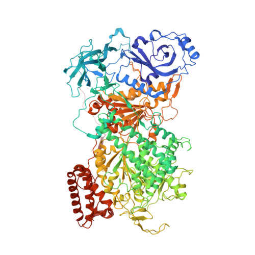
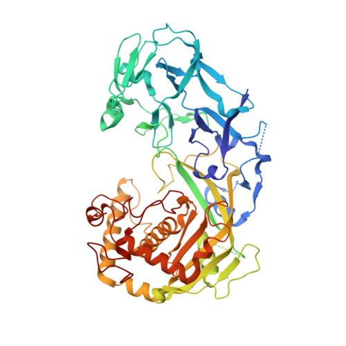
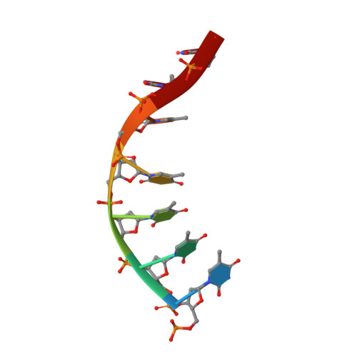
Structural basis for broad anti-phage immunity by DISARM.

Bravo, J.P.K.Aparicio-Maldonado, C.Nobrega, F.L.Brouns, S.J.J.Taylor, D.W.

(2022) Nat Commun 13: 2987-2987

  • DOI: https://doi.org/10.1038/s41467-022-30673-1
  • Primary Citation Related Structures: 
    7S9V, 7S9W

  • PubMed Abstract: 

    In the evolutionary arms race against phage, bacteria have assembled a diverse arsenal of antiviral immune strategies. While the recently discovered DISARM (Defense Island System Associated with Restriction-Modification) systems can provide protection against a wide range of phage, the molecular mechanisms that underpin broad antiviral targeting but avoiding autoimmunity remain enigmatic. Here, we report cryo-EM structures of the core DISARM complex, DrmAB, both alone and in complex with an unmethylated phage DNA mimetic. These structures reveal that DrmAB core complex is autoinhibited by a trigger loop (TL) within DrmA and binding to DNA substrates containing a 5' overhang dislodges the TL, initiating a long-range structural rearrangement for DrmAB activation. Together with structure-guided in vivo studies, our work provides insights into the mechanism of phage DNA recognition and specific activation of this widespread antiviral defense system.


  • Organizational Affiliation
    • Department of Molecular Biosciences, University of Texas at Austin, Austin, TX, 78712, USA.

Macromolecule Content 

  • Total Structure Weight: 219.16 kDa 
  • Atom Count: 14,052 
  • Modeled Residue Count: 1,774 
  • Deposited Residue Count: 1,951 
  • Unique protein chains: 2
  • Unique nucleic acid chains: 1

Macromolecules


Find similar proteins by:|  3D Structure
Entity ID: 1
MoleculeChains  Sequence LengthOrganismDetailsImage
DrmA1,325SerratiaMutation(s): 0 
UniProt
Find proteins for A0A979HMQ0 (Serratia)
Explore A0A979HMQ0 
Go to UniProtKB:  A0A979HMQ0
Entity Groups
Sequence Clusters30% Identity50% Identity70% Identity90% Identity95% Identity100% Identity
UniProt GroupA0A979HMQ0
Sequence Annotations
Expand
Reference Sequence
Find similar proteins by:|  3D Structure
Entity ID: 2
MoleculeChains  Sequence LengthOrganismDetailsImage
DrmB619SerratiaMutation(s): 0 
UniProt
Find proteins for A0A979HMQ1 (Serratia)
Explore A0A979HMQ1 
Go to UniProtKB:  A0A979HMQ1
Entity Groups
Sequence Clusters30% Identity50% Identity70% Identity90% Identity95% Identity100% Identity
UniProt GroupA0A979HMQ1
Sequence Annotations
Expand
Reference Sequence
Find similar nucleic acids by:  Sequence
Entity ID: 3
MoleculeChains LengthOrganismImage
DNA (5'-D(P*TP*TP*TP*TP*TP*TP*T)-3')7synthetic construct
Sequence Annotations
Expand
Reference Sequence

Small Molecules

Ligands 1 Unique
IDChains Name / Formula / InChI Key2D Diagram3D Interactions
ADP
(Subject of Investigation/LOI)

Query on ADP


Download:Ideal Coordinates CCD File
D [auth A]ADENOSINE-5'-DIPHOSPHATE
C10 H15 N5 O10 P2
XTWYTFMLZFPYCI-KQYNXXCUSA-N

Experimental Data & Validation

Experimental Data

  • Method: ELECTRON MICROSCOPY
  • Resolution: 3.40 Å
  • Aggregation State: PARTICLE 
  • Reconstruction Method: SINGLE PARTICLE 
EM Software:
TaskSoftware PackageVersion
MODEL REFINEMENTPHENIX

Structure Validation

View Full Validation Report


Entry History 

& Funding Information

Deposition Data


Funding OrganizationLocationGrant Number
Welch FoundationUnited StatesF-1938

Revision History  (Full details and data files)

  • Version 1.0: 2022-06-15
    Type: Initial release
  • Version 1.1: 2024-06-05
    Changes: Data collection
  • Version 1.2: 2025-05-14
    Changes: Data collection, Structure summary