


Crystal Structure of a 8-amino-7-oxononanoate synthase/2-amino-3-ketobutyrate coenzyme A ligase from Mycobacterium smegmatis
Abendroth, J., Davies, D.R., Sankaran, B., Lorimer, D.D., Horanyi, P.S., Edwards, T.E.To be published.
Experimental Data Snapshot
Starting Model: experimental
View more details
wwPDB Validation 3D Report Full Report

Entity ID: 1 | |||||
|---|---|---|---|---|---|
| Molecule | Chains | Sequence Length | Organism | Details | Image |
| 8-amino-7-oxononanoate synthase | 404 | Mycolicibacterium smegmatis MC2 155 | Mutation(s): 0 Gene Names: bioF, MSMEG_3189, MSMEI_3107 EC: 2.3.1.47 | 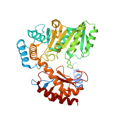 | |
UniProt | |||||
Entity Groups | |||||
| Sequence Clusters | 30% Identity50% Identity70% Identity90% Identity95% Identity100% Identity | ||||
| UniProt Group | A0QX65 | ||||
Sequence AnnotationsExpand | |||||
Reference Sequence | |||||
| Length ( Å ) | Angle ( ˚ ) |
|---|---|
| a = 60.71 | α = 90 |
| b = 108.94 | β = 94.651 |
| c = 108.34 | γ = 90 |
| Software Name | Purpose |
|---|---|
| XDS | data reduction |
| XSCALE | data scaling |
| PHENIX | refinement |
| PDB_EXTRACT | data extraction |
| MoRDa | phasing |
| Coot | model building |


