7S4M | pdb_00007s4m

CryoEM structure of Methylocystis sp. str. Rockwell pMMO in a POPC nanodisc at 2.42 Angstrom resolution


Experimental Data Snapshot

  • Method: ELECTRON MICROSCOPY
  • Resolution: 2.42 Å
  • Aggregation State: PARTICLE 
  • Reconstruction Method: SINGLE PARTICLE 

wwPDB Validation 3D Report Full Report

Validation slider image for 7S4M

This is version 1.1 of the entry. See complete history

Literature

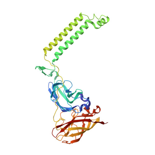
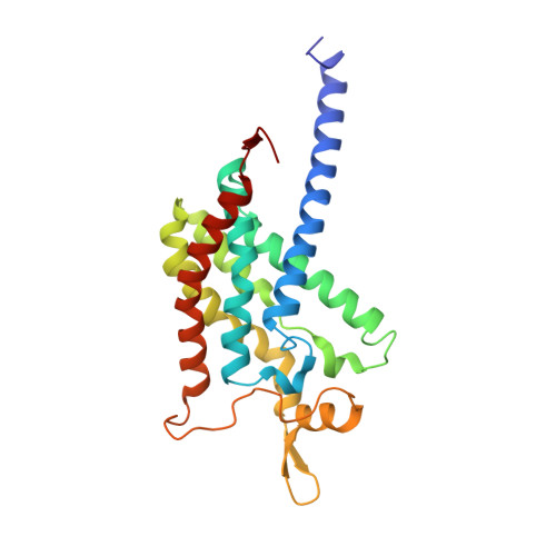
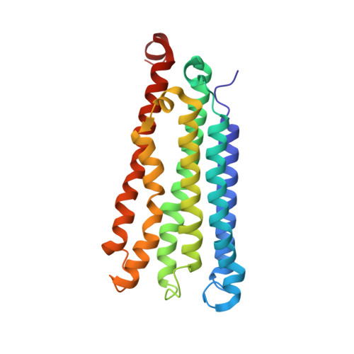
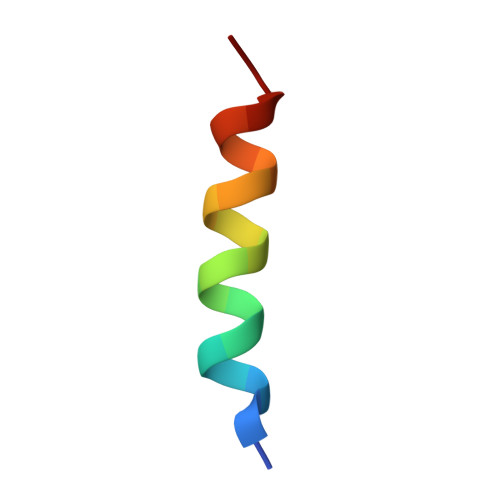
Recovery of particulate methane monooxygenase structure and activity in a lipid bilayer.

Koo, C.W.Tucci, F.J.He, Y.Rosenzweig, A.C.

(2022) Science 375: 1287-1291

  • DOI: https://doi.org/10.1126/science.abm3282
  • Primary Citation Related Structures: 
    7S4H, 7S4I, 7S4J, 7S4K, 7S4L, 7S4M, 7T4O, 7T4P

  • PubMed Abstract: 

    Bacterial methane oxidation using the enzyme particulate methane monooxygenase (pMMO) contributes to the removal of environmental methane, a potent greenhouse gas. Crystal structures determined using inactive, detergent-solubilized pMMO lack several conserved regions neighboring the proposed active site. We show that reconstituting pMMO in nanodiscs with lipids extracted from the native organism restores methane oxidation activity. Multiple nanodisc-embedded pMMO structures determined by cryo-electron microscopy to 2.14- to 2.46-angstrom resolution reveal the structure of pMMO in a lipid environment. The resulting model includes stabilizing lipids, regions of the PmoA and PmoC subunits not observed in prior structures, and a previously undetected copper-binding site in the PmoC subunit with an adjacent hydrophobic cavity. These structures provide a revised framework for understanding and engineering pMMO function.


  • Organizational Affiliation
    • Department of Molecular Biosciences, Northwestern University, Evanston, IL 60208, USA.

Macromolecule Content 

  • Total Structure Weight: 307.63 kDa 
  • Atom Count: 21,729 
  • Modeled Residue Count: 2,676 
  • Deposited Residue Count: 2,676 
  • Unique protein chains: 4

Macromolecules

Find similar proteins by:|  3D Structure
Entity ID: 1
MoleculeChains  Sequence LengthOrganismDetailsImage
Particulate methane monooxygenase alpha subunitA,
K [auth E],
L [auth I]
388Methylocystis sp. ATCC 49242Mutation(s): 0 
Find similar proteins by:|  3D Structure
Entity ID: 2
MoleculeChains  Sequence LengthOrganismDetailsImage
Particulate methane monooxygenase beta subunitB [auth F],
C [auth B],
D [auth J]
244Methylocystis sp. ATCC 49242Mutation(s): 0 
UniProt
Find proteins for A0ACD6B8G4 (Methylocystis sp. ATCC 49242)
Explore A0ACD6B8G4 
Go to UniProtKB:  A0ACD6B8G4
Entity Groups
Sequence Clusters30% Identity50% Identity70% Identity90% Identity95% Identity100% Identity
UniProt GroupA0ACD6B8G4
Sequence Annotations
Expand
Reference Sequence
Find similar proteins by:|  3D Structure
Entity ID: 3
MoleculeChains  Sequence LengthOrganismDetailsImage
Ammonia monooxygenase/methane monooxygenase, subunit C family proteinE [auth G],
F [auth C],
H [auth K]
241Methylocystis sp. ATCC 49242Mutation(s): 0 
Find similar proteins by:  Sequence   |   3D Structure  
Entity ID: 4
MoleculeChains  Sequence LengthOrganismDetailsImage
Unidentified HelixG [auth D],
I [auth N],
J [auth H]
19Methylocystis sp. ATCC 49242Mutation(s): 0 

Small Molecules

Ligands 4 Unique
IDChains Name / Formula / InChI Key2D Diagram3D Interactions
HXG
(Subject of Investigation/LOI)

Query on HXG


Download:Ideal Coordinates CCD File
CA [auth C]
EA [auth C]
HA [auth K]
JA [auth K]
N [auth F]
CA [auth C],
EA [auth C],
HA [auth K],
JA [auth K],
N [auth F],
O [auth F],
Q [auth B],
R [auth B],
T [auth J],
U [auth J],
X [auth G],
Z [auth G]
1,2-dihexanoyl-sn-glycero-3-phosphocholine
C20 H41 N O8 P
DVZARZBAWHITHR-GOSISDBHSA-O
D12
(Subject of Investigation/LOI)

Query on D12


Download:Ideal Coordinates CCD File
DA [auth C],
IA [auth K],
Y [auth G]
DODECANE
C12 H26
SNRUBQQJIBEYMU-UHFFFAOYSA-N
D10
(Subject of Investigation/LOI)

Query on D10


Download:Ideal Coordinates CCD File
AA [auth G]
FA [auth C]
KA [auth K]
P [auth F]
S [auth B]
AA [auth G],
FA [auth C],
KA [auth K],
P [auth F],
S [auth B],
V [auth J]
DECANE
C10 H22
DIOQZVSQGTUSAI-UHFFFAOYSA-N
CU
(Subject of Investigation/LOI)

Query on CU


Download:Ideal Coordinates CCD File
BA [auth C]
GA [auth K]
LA [auth E]
M [auth A]
MA [auth I]
BA [auth C],
GA [auth K],
LA [auth E],
M [auth A],
MA [auth I],
W [auth G]
COPPER (II) ION
Cu
JPVYNHNXODAKFH-UHFFFAOYSA-N

Experimental Data & Validation

Experimental Data

  • Method: ELECTRON MICROSCOPY
  • Resolution: 2.42 Å
  • Aggregation State: PARTICLE 
  • Reconstruction Method: SINGLE PARTICLE 

Structure Validation

View Full Validation Report


Entry History 

& Funding Information

Deposition Data


Funding OrganizationLocationGrant Number
National Institutes of Health/National Institute of General Medical Sciences (NIH/NIGMS)United States--

Revision History  (Full details and data files)

  • Version 1.0: 2022-03-30
    Type: Initial release
  • Version 1.1: 2024-06-05
    Changes: Data collection