Molecular Dynamics Simulations Guide Chimeragenesis and Engineered Control of Chemoselectivity in Diketopiperazine Dimerases.
Shende, V.V., Harris, N.R., Sanders, J.N., Newmister, S.A., Khatri, Y., Movassaghi, M., Houk, K.N., Sherman, D.H.(2023) Angew Chem Int Ed Engl 62: e202210254-e202210254
- PubMed: 36610039 Search on PubMedSearch on PubMed Central
- DOI: https://doi.org/10.1002/anie.202210254
- Primary Citation Related Structures:
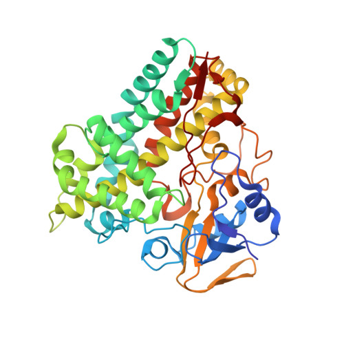
7S3J, 7S3T - PubMed Abstract:
In the biosynthesis of the tryptophan-linked dimeric diketopiperazines (DKPs), cytochromes P450 selectively couple DKP monomers to generate a variety of intricate and isomeric frameworks. To determine the molecular basis for selectivity of these biocatalysts we obtained a high-resolution crystal structure of selective Csp 2 -N bond forming dimerase, AspB. Overlay of the AspB structure onto C-C and C-N bond forming homolog NzeB revealed no significant structural variance to explain their divergent chemoselectivities. Molecular dynamics (MD) simulations identified a region of NzeB with increased conformational flexibility relative to AspB, and interchange of this region along with a single active site mutation led to a variant that catalyzes exclusive C-N bond formation. MD simulations also suggest that intermolecular C-C or C-N bond formation results from a change in mechanism, supported experimentally through use of a substrate mimic.
- Life Sciences Institute, University of Michigan, Ann Arbor, MÌ 48109, USA.
Organizational Affiliation: