


Self-association configures the NAD + -binding site of plant NLR TIR domains
Burdett, H., Hu, X., Rank, M.X., Maruta, N., Kobe, B.(2021) bioRxiv
Experimental Data Snapshot
Starting Model: experimental
View more details
(2021) bioRxiv
Entity ID: 1 | |||||
|---|---|---|---|---|---|
| Molecule | Chains | Sequence Length | Organism | Details | Image |
| Disease resistance protein RUN1 | 179 | Vitis rotundifolia | Mutation(s): 1 Gene Names: RUN1 EC: 3.2.2.6 (PDB Primary Data), 3.2.2 (PDB Primary Data) | 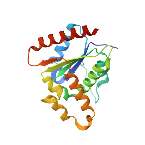 | |
UniProt | |||||
Entity Groups | |||||
| Sequence Clusters | 30% Identity50% Identity70% Identity90% Identity95% Identity100% Identity | ||||
| UniProt Group | V9M398 | ||||
Sequence AnnotationsExpand | |||||
Reference Sequence | |||||
| Ligands 2 Unique | |||||
|---|---|---|---|---|---|
| ID | Chains | Name / Formula / InChI Key | 2D Diagram | 3D Interactions | |
| NAD (Subject of Investigation/LOI) Download:Ideal Coordinates CCD File | E [auth A], K [auth B], Q [auth C], V [auth D] | NICOTINAMIDE-ADENINE-DINUCLEOTIDE C21 H27 N7 O14 P2 BAWFJGJZGIEFAR-NNYOXOHSSA-N |  | ||
| SO4 Download:Ideal Coordinates CCD File | F [auth A] G [auth A] H [auth A] I [auth A] J [auth A] | SULFATE ION O4 S QAOWNCQODCNURD-UHFFFAOYSA-L |  | ||
| Length ( Å ) | Angle ( ˚ ) |
|---|---|
| a = 79.743 | α = 90 |
| b = 116.644 | β = 90 |
| c = 122.22 | γ = 90 |
| Software Name | Purpose |
|---|---|
| PHENIX | refinement |
| XDS | data reduction |
| Aimless | data scaling |
| PHASER | phasing |
| Funding Organization | Location | Grant Number |
|---|---|---|
| Australian Research Council (ARC) | Australia | DP160102244 |
| Australian Research Council (ARC) | Australia | DP190102526 |


