7S2S | pdb_00007s2s

nanobody bound to Interleukin-2Rbeta


Experimental Data Snapshot

  • Method: X-RAY DIFFRACTION
  • Resolution: 1.93 Å
  • R-Value Free: 
    0.247 (Depositor), 0.248 (DCC) 
  • R-Value Work: 
    0.213 (Depositor), 0.214 (DCC) 
  • R-Value Observed: 
    0.214 (Depositor) 

Starting Models: experimental
View more details

wwPDB Validation 3D Report Full Report

Validation slider image for 7S2S

This is version 1.3 of the entry. See complete history

Literature

Facile discovery of surrogate cytokine agonists.

Yen, M.Ren, J.Liu, Q.Glassman, C.R.Sheahan, T.P.Picton, L.K.Moreira, F.R.Rustagi, A.Jude, K.M.Zhao, X.Blish, C.A.Baric, R.S.Su, L.L.Garcia, K.C.

(2022) Cell 185: 1414

  • DOI: https://doi.org/10.1016/j.cell.2022.02.025
  • Primary Citation Related Structures: 
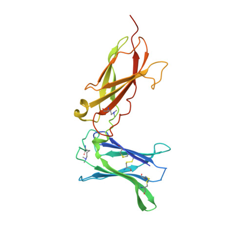
    7S2R, 7S2S

  • PubMed Abstract: 

    Cytokines are powerful immune modulators that initiate signaling through receptor dimerization, but natural cytokines have structural limitations as therapeutics. We present a strategy to discover cytokine surrogate agonists by using modular ligands that exploit induced proximity and receptor dimer geometry as pharmacological metrics amenable to high-throughput screening. Using VHH and scFv to human interleukin-2/15, type-I interferon, and interleukin-10 receptors, we generated combinatorial matrices of single-chain bispecific ligands that exhibited diverse spectrums of functional activities, including potent inhibition of SARS-CoV-2 by surrogate interferons. Crystal structures of IL-2R:VHH complexes revealed that variation in receptor dimer geometries resulted in functionally diverse signaling outputs. This modular platform enabled engineering of surrogate ligands that compelled assembly of an IL-2R/IL-10R heterodimer, which does not naturally exist, that signaled through pSTAT5 on T and natural killer (NK) cells. This "cytokine med-chem" approach, rooted in principles of induced proximity, is generalizable for discovery of diversified agonists for many ligand-receptor systems.


  • Organizational Affiliation
    • Departments of Molecular and Cellular Physiology, and Structural Biology, Stanford University School of Medicine, Stanford, CA 94305, USA; Howard Hughes Medical Institute, Stanford University School of Medicine, Stanford, CA 94305, USA.

Macromolecule Content 

  • Total Structure Weight: 88.27 kDa 
  • Atom Count: 5,660 
  • Modeled Residue Count: 657 
  • Deposited Residue Count: 740 
  • Unique protein chains: 2

Macromolecules

Find similar proteins by:|  3D Structure
Entity ID: 1
MoleculeChains  Sequence LengthOrganismDetailsImage
IL2Rb-binding nanobodyA [auth C],
C [auth A]
154Camelus bactrianusMutation(s): 0 
Find similar proteins by:|  3D Structure
Entity ID: 2
MoleculeChains  Sequence LengthOrganismDetailsImage
Interleukin-2 receptor subunit beta
B, D
216Homo sapiensMutation(s): 0 
Gene Names: IL2RBIL15RB
UniProt & NIH Common Fund Data Resources
Find proteins for P14784 (Homo sapiens)
Explore P14784 
Go to UniProtKB:  P14784
PHAROS:  P14784
GTEx:  ENSG00000100385 
Entity Groups
Sequence Clusters30% Identity50% Identity70% Identity90% Identity95% Identity100% Identity
UniProt GroupP14784
Glycosylation
Glycosylation Sites: 3Go to GlyGen: P14784-1
Sequence Annotations
Expand
Reference Sequence

Oligosaccharides

Help  
Entity ID: 3
MoleculeChains Length2D Diagram GlycosylationD Interactions
2-acetamido-2-deoxy-beta-D-glucopyranose-(1-4)-[alpha-L-fucopyranose-(1-6)]2-acetamido-2-deoxy-beta-D-glucopyranose
E, F, H, I
3N-Glycosylation
Glycosylation Resources
GlyTouCan: G21290RB
GlyCosmos: G21290RB
GlyGen: G21290RB
Entity ID: 4
MoleculeChains Length2D Diagram GlycosylationD Interactions
beta-D-mannopyranose-(1-4)-2-acetamido-2-deoxy-beta-D-glucopyranose-(1-4)-2-acetamido-2-deoxy-beta-D-glucopyranose
G, J
3N-Glycosylation
Glycosylation Resources
GlyTouCan: G15407YE
GlyCosmos: G15407YE
GlyGen: G15407YE

Small Molecules

Ligands 1 Unique
IDChains Name / Formula / InChI Key2D Diagram3D Interactions
SO4

Query on SO4


Download:Ideal Coordinates CCD File
K [auth B]
L [auth B]
M [auth B]
N [auth B]
O [auth A]
K [auth B],
L [auth B],
M [auth B],
N [auth B],
O [auth A],
P [auth D],
Q [auth D],
R [auth D],
S [auth D],
T [auth D],
U [auth D],
V [auth D],
W [auth D]
SULFATE ION
O4 S
QAOWNCQODCNURD-UHFFFAOYSA-L
Modified Residues  1 Unique
IDChains TypeFormula2D DiagramParent
MLY
Query on MLY
A [auth C],
C [auth A]
L-PEPTIDE LINKINGC8 H18 N2 O2LYS

Experimental Data & Validation

Experimental Data

  • Method: X-RAY DIFFRACTION
  • Resolution: 1.93 Å
  • R-Value Free:  0.247 (Depositor), 0.248 (DCC) 
  • R-Value Work:  0.213 (Depositor), 0.214 (DCC) 
  • R-Value Observed: 0.214 (Depositor) 
Space Group: P 1
Unit Cell:
Length ( Å )Angle ( ˚ )
a = 42.698α = 99.566
b = 69.505β = 99.854
c = 91.721γ = 90.191
Software Package:
Software NamePurpose
PHENIXrefinement
XDSdata reduction
Aimlessdata scaling
PHASERphasing
PDB_EXTRACTdata extraction

Structure Validation

View Full Validation Report


Entry History 

& Funding Information

Deposition Data


Funding OrganizationLocationGrant Number
National Institutes of Health/National Institute Of Allergy and Infectious Diseases (NIH/NIAID)United StatesR37-AI051321

Revision History  (Full details and data files)

  • Version 1.0: 2022-03-30
    Type: Initial release
  • Version 1.1: 2022-04-06
    Changes: Database references
  • Version 1.2: 2022-04-27
    Changes: Database references
  • Version 1.3: 2023-10-18
    Changes: Data collection, Refinement description