Tryptophan mutations in G3BP1 tune the stability of a cellular signaling hub by weakening transient interactions with Caprin1 and USP10.
Sheehan, C.T., Hampton, T.H., Madden, D.R.(2022) J Biological Chem 298: 102552-102552
- PubMed: 36183834 Search on PubMedSearch on PubMed Central
- DOI: https://doi.org/10.1016/j.jbc.2022.102552
- Primary Citation Related Structures:
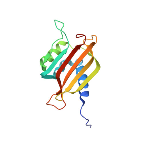
7S17 - PubMed Abstract:
Intrinsically disordered proteins (IDPs) often coordinate transient interactions with multiple proteins to mediate complex signals within large protein networks. Among these, the IDP hub protein G3BP1 can form complexes with cytoplasmic phosphoprotein Caprin1 and ubiquitin peptidase USP10; the resulting control of USP10 activity contributes to a pathogenic virulence system that targets endocytic recycling of the ion channel CFTR. However, while the identities of protein interactors are known for many IDP hub proteins, the relationship between pairwise affinities and the extent of protein recruitment and activity is not well understood. Here, we describe in vitro analysis of these G3BP1 affinities and show tryptophan substitutions of specific G3BP1 residues reduce its affinity for both USP10 and Caprin1. We show that these same mutations reduce the stability of complexes between the full-length proteins, suggesting that copurification can serve as a surrogate measure of interaction strength. The crystal structure of G3BP1 TripleW (F15W/F33W/F124W) mutant reveals a clear reorientation of the side chain of W33, creating a steric clash with USP10 and Caprin1. Furthermore, an amino-acid scan of USP10 and Caprin1 peptides reveals similarities and differences in the ability to substitute residues in the core motifs as well as specific substitutions with the potential to create higher affinity peptides. Taken together, these data show that small changes in component binding affinities can have significant effects on the composition of cellular interaction hubs. These specific protein mutations can be harnessed to manipulate complex protein networks, informing future investigations into roles of these networks in cellular processes.
- Department of Biochemistry and Cell Biology, Geisel School of Medicine at Dartmouth, Hanover, New Hampshire, USA.
Organizational Affiliation: