Mutation of the conserved Asp-Asp pair impairs the structure, function, and inhibition of CTX-M Class A beta-lactamase.
Kemp, M.T., Nichols, D.A., Zhang, X., Defrees, K., Na, I., Renslo, A.R., Chen, Y.(2021) FEBS Lett 595: 2981-2994
- PubMed: 34704263 Search on PubMedSearch on PubMed Central
- DOI: https://doi.org/10.1002/1873-3468.14215
- Primary Citation Related Structures:
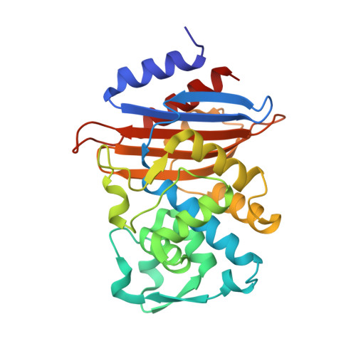
7S0V - PubMed Abstract:
The Asp233-Asp246 pair is highly conserved in Class A β-lactamases, which hydrolyze β-lactam antibiotics. Here, we characterize its function using CTX-M-14 β-lactamase. The D233N mutant displayed decreased activity that is substrate-dependent, with reductions in k cat /K m ranging from 20% for nitrocefin to 6-fold for cefotaxime. In comparison, the mutation reduced the binding of a known reversible inhibitor by 10-fold. The mutant structures showed movement of the 213-219 loop and the loss of the Thr216-Thr235 hydrogen bond, which was restored by inhibitor binding. Mutagenesis of Thr216 further highlighted its contribution to CTX-M activity. These results demonstrate the importance of the aspartate pair to CTX-M hydrolysis of substrates with bulky side chains, while suggesting increased protein flexibility as a means to evolve drug resistance.
- Department of Molecular Medicine, University of South Florida College of Medicine, Tampa, FL, USA.
Organizational Affiliation: