7S0K

HAP2 from Cyanidioschyzon merolae


Experimental Data Snapshot

  • Method: X-RAY DIFFRACTION
  • Resolution: 2.30 Å
  • R-Value Free: 0.264 
  • R-Value Work: 0.223 
  • R-Value Observed: 0.226 

wwPDB Validation   3D Report Full Report


Ligand Structure Quality Assessment 


This is version 1.2 of the entry. See complete history


Literature

Monomeric prefusion structure of an extremophile gamete fusogen and stepwise formation of the postfusion trimeric state.

Feng, J.Dong, X.Su, Y.Lu, C.Springer, T.A.

(2022) Nat Commun 13: 4064-4064

  • DOI: https://doi.org/10.1038/s41467-022-31744-z
  • Primary Citation of Related Structures:  
    7S0K

  • PubMed Abstract: 

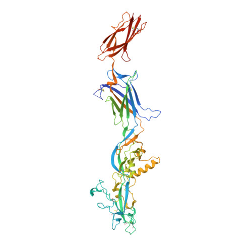
    Here, we study the gamete fusogen HAP2 from Cyanidioschyzon merolae (Cyani), an extremophile red algae that grows at acidic pH at 45 °C. HAP2 has a trimeric postfusion structure with similarity to viral class II fusion proteins, but its prefusion structure has been elusive. The crystal structure of a monomeric prefusion state of Cyani HAP2 shows it is highly extended with three domains in the order D2, D1, and D3. Three hydrophobic fusion loops at the tip of D2 are each required for postfusion state formation. We followed by negative stain electron microscopy steps in the process of detergent micelle-stimulated postfusion state formation. In an intermediate state, two or three linear HAP2 monomers associate at the end of D2 bearing its fusion loops. Subsequently, D2 and D1 line the core of a trimer and D3 folds back over the exterior of D1 and D2. D3 is not required for formation of intermediate or postfusion-like states.


  • Organizational Affiliation

    Program in Cellular and Molecular Medicine, Department of Pediatrics, Boston Children's Hospital, Boston, MA, USA.


Macromolecules
Find similar proteins by:  (by identity cutoff)  |  3D Structure
Entity ID: 1
MoleculeChains Sequence LengthOrganismDetailsImage
HAP2-GCS1 domain-containing protein545Cyanidioschyzon merolaeMutation(s): 0 
Gene Names: CYME_CMK076C
UniProt
Find proteins for M1V8A6 (Cyanidioschyzon merolae (strain NIES-3377 / 10D))
Explore M1V8A6 
Go to UniProtKB:  M1V8A6
Entity Groups  
Sequence Clusters30% Identity50% Identity70% Identity90% Identity95% Identity100% Identity
UniProt GroupM1V8A6
Sequence Annotations
Expand
  • Reference Sequence
Oligosaccharides

Help

Entity ID: 2
MoleculeChains Length2D Diagram Glycosylation3D Interactions
alpha-D-mannopyranose-(1-3)-beta-D-mannopyranose-(1-3)-beta-D-mannopyranose-(1-4)-2-acetamido-2-deoxy-beta-D-glucopyranose-(1-4)-2-acetamido-2-deoxy-beta-D-glucopyranose
B
5N-Glycosylation
Glycosylation Resources
GlyTouCan:  G52322TF
GlyCosmos:  G52322TF
GlyGen:  G52322TF
Experimental Data & Validation

Experimental Data

  • Method: X-RAY DIFFRACTION
  • Resolution: 2.30 Å
  • R-Value Free: 0.264 
  • R-Value Work: 0.223 
  • R-Value Observed: 0.226 
  • Space Group: C 2 2 21
Unit Cell:
Length ( Å )Angle ( ˚ )
a = 102.96α = 90
b = 197.01β = 90
c = 74.18γ = 90
Software Package:
Software NamePurpose
PHENIXrefinement
XSCALEdata scaling
PDB_EXTRACTdata extraction
Cootmodel building
XSCALEdata scaling
PHASERphasing

Structure Validation

View Full Validation Report


Ligand Structure Quality Assessment 


Entry History & Funding Information

Deposition Data


Funding OrganizationLocationGrant Number
National Institutes of Health/Eunice Kennedy Shriver National Institute of Child Health & Human Development (NIH/NICHD)United States--

Revision History  (Full details and data files)

  • Version 1.0: 2022-07-06
    Type: Initial release
  • Version 1.1: 2022-07-27
    Changes: Database references
  • Version 1.2: 2023-10-18
    Changes: Data collection, Refinement description