7S00

X-ray structure of the phage AR9 non-virion RNA polymerase core


Experimental Data Snapshot

  • Method: X-RAY DIFFRACTION
  • Resolution: 3.30 Å
  • R-Value Free: 0.258 
  • R-Value Work: 0.219 
  • R-Value Observed: 0.221 

wwPDB Validation   3D Report Full Report


This is version 1.1 of the entry. See complete history


Literature

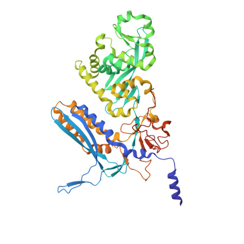
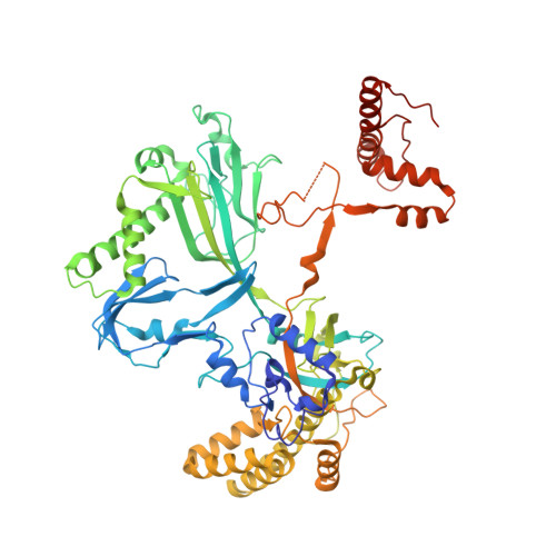
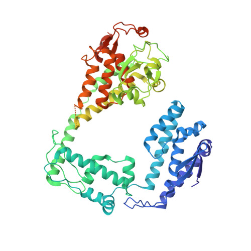
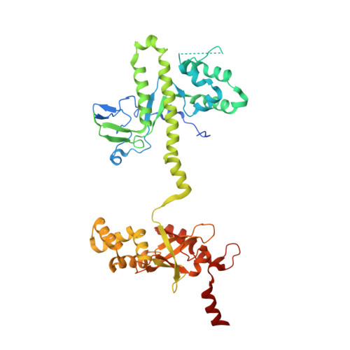
Structural basis of template strand deoxyuridine promoter recognition by a viral RNA polymerase.

Fraser, A.Sokolova, M.L.Drobysheva, A.V.Gordeeva, J.V.Borukhov, S.Jumper, J.Severinov, K.V.Leiman, P.G.

(2022) Nat Commun 13: 3526-3526

  • DOI: https://doi.org/10.1038/s41467-022-31214-6
  • Primary Citation of Related Structures:  
    7S00, 7S01, 7UM0, 7UM1

  • PubMed Abstract: 

    Recognition of promoters in bacterial RNA polymerases (RNAPs) is controlled by sigma subunits. The key sequence motif recognized by the sigma, the -10 promoter element, is located in the non-template strand of the double-stranded DNA molecule ~10 nucleotides upstream of the transcription start site. Here, we explain the mechanism by which the phage AR9 non-virion RNAP (nvRNAP), a bacterial RNAP homolog, recognizes the -10 element of its deoxyuridine-containing promoter in the template strand. The AR9 sigma-like subunit, the nvRNAP enzyme core, and the template strand together form two nucleotide base-accepting pockets whose shapes dictate the requirement for the conserved deoxyuridines. A single amino acid substitution in the AR9 sigma-like subunit allows one of these pockets to accept a thymine thus expanding the promoter consensus. Our work demonstrates the extent to which viruses can evolve host-derived multisubunit enzymes to make transcription of their own genes independent of the host.


  • Organizational Affiliation

    Department of Biochemistry and Molecular Biology, Sealy Center for Structural Biology and Molecular Biophysics, University of Texas Medical Branch, Galveston, TX, 77555-0647, USA.


Macromolecules
Find similar proteins by:  (by identity cutoff)  |  3D Structure
Entity ID: 1
MoleculeChains Sequence LengthOrganismDetailsImage
DNA-directed RNA polymerase beta subunitA [auth c],
E [auth e]
496Bacillus phage AR9Mutation(s): 0 
Gene Names: AR9_g105
UniProt
Find proteins for A0A172JI16 (Bacillus phage AR9)
Explore A0A172JI16 
Go to UniProtKB:  A0A172JI16
Entity Groups  
Sequence Clusters30% Identity50% Identity70% Identity90% Identity95% Identity100% Identity
UniProt GroupA0A172JI16
Sequence Annotations
Expand
  • Reference Sequence
Find similar proteins by:  (by identity cutoff)  |  3D Structure
Entity ID: 2
MoleculeChains Sequence LengthOrganismDetailsImage
DNA-directed RNA polymeraseB [auth C],
F [auth E]
665Bacillus phage AR9Mutation(s): 0 
Gene Names: AR9_g089
EC: 2.7.7.6
UniProt
Find proteins for A0A172JHZ2 (Bacillus phage AR9)
Explore A0A172JHZ2 
Go to UniProtKB:  A0A172JHZ2
Entity Groups  
Sequence Clusters30% Identity50% Identity70% Identity90% Identity95% Identity100% Identity
UniProt GroupA0A172JHZ2
Sequence Annotations
Expand
  • Reference Sequence
Find similar proteins by:  (by identity cutoff)  |  3D Structure
Entity ID: 3
MoleculeChains Sequence LengthOrganismDetailsImage
DNA-directed RNA polymeraseC [auth D],
G [auth F]
631Bacillus phage AR9Mutation(s): 0 
Gene Names: AR9_g154
EC: 2.7.7.6
UniProt
Find proteins for A0A172JI62 (Bacillus phage AR9)
Explore A0A172JI62 
Go to UniProtKB:  A0A172JI62
Entity Groups  
Sequence Clusters30% Identity50% Identity70% Identity90% Identity95% Identity100% Identity
UniProt GroupA0A172JI62
Sequence Annotations
Expand
  • Reference Sequence
Find similar proteins by:  (by identity cutoff)  |  3D Structure
Entity ID: 4
MoleculeChains Sequence LengthOrganismDetailsImage
DNA-directed RNA polymerase beta' subunitD [auth d],
H [auth f]
448Bacillus phage AR9Mutation(s): 0 
Gene Names: AR9_g270
UniProt
Find proteins for A0A172JIH0 (Bacillus phage AR9)
Explore A0A172JIH0 
Go to UniProtKB:  A0A172JIH0
Entity Groups  
Sequence Clusters30% Identity50% Identity70% Identity90% Identity95% Identity100% Identity
UniProt GroupA0A172JIH0
Sequence Annotations
Expand
  • Reference Sequence
Experimental Data & Validation

Experimental Data

  • Method: X-RAY DIFFRACTION
  • Resolution: 3.30 Å
  • R-Value Free: 0.258 
  • R-Value Work: 0.219 
  • R-Value Observed: 0.221 
  • Space Group: P 21 21 21
Unit Cell:
Length ( Å )Angle ( ˚ )
a = 112.858α = 90
b = 166.27β = 90
c = 307.215γ = 90
Software Package:
Software NamePurpose
Cootmodel building
PHENIXrefinement
XDSdata reduction
XDSdata scaling
PHASERphasing

Structure Validation

View Full Validation Report


Entry History & Funding Information

Deposition Data


Funding OrganizationLocationGrant Number
Not fundedFunded by UTMB

Revision History  (Full details and data files)

  • Version 1.0: 2022-07-06
    Type: Initial release
  • Version 1.1: 2023-10-18
    Changes: Data collection, Refinement description