7RYZ

Structure of the complex of LBD-TMD part of AMPA receptor GluA2 with auxiliary subunit GSG1L bound to agonist quisqualate


Experimental Data Snapshot

  • Method: ELECTRON MICROSCOPY
  • Resolution: 4.15 Å
  • Aggregation State: PARTICLE 
  • Reconstruction Method: SINGLE PARTICLE 

wwPDB Validation   3D Report Full Report


This is version 1.1 of the entry. See complete history


Literature

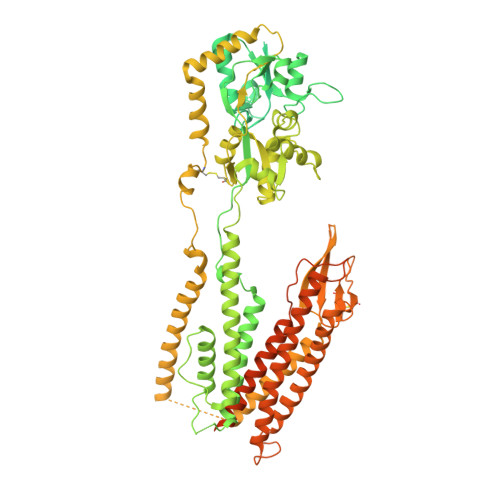
Structure and desensitization of AMPA receptor complexes with type II TARP gamma 5 and GSG1L.

Klykov, O.Gangwar, S.P.Yelshanskaya, M.V.Yen, L.Sobolevsky, A.I.

(2021) Mol Cell 81: 4771-4783.e7

  • DOI: https://doi.org/10.1016/j.molcel.2021.09.030
  • Primary Citation of Related Structures:  
    7RYY, 7RYZ, 7RZ4, 7RZ5, 7RZ6, 7RZ7, 7RZ8, 7RZ9, 7RZA

  • PubMed Abstract: 

    AMPA receptors (AMPARs) mediate the majority of excitatory neurotransmission. Their surface expression, trafficking, gating, and pharmacology are regulated by auxiliary subunits. Of the two types of TARP auxiliary subunits, type I TARPs assume activating roles, while type II TARPs serve suppressive functions. We present cryo-EM structures of GluA2 AMPAR in complex with type II TARP γ5, which reduces steady-state currents, increases single-channel conductance, and slows recovery from desensitization. Regulation of AMPAR function depends on its ligand-binding domain (LBD) interaction with the γ5 head domain. GluA2-γ5 complex shows maximum stoichiometry of two TARPs per AMPAR tetramer, being different from type I TARPs but reminiscent of the auxiliary subunit GSG1L. Desensitization of both GluA2-GSG1L and GluA2-γ5 complexes is accompanied by rupture of LBD dimer interface, while GluA2-γ5 but not GluA2-GSG1L LBD dimers remain two-fold symmetric. Different structural architectures and desensitization mechanisms of complexes with auxiliary subunits endow AMPARs with broad functional capabilities.


  • Organizational Affiliation

    Department of Biochemistry and Molecular Biophysics, Columbia University, 650 West 168th Street, New York, NY 10032, USA.


Macromolecules
Find similar proteins by:  (by identity cutoff)  |  3D Structure
Entity ID: 1
MoleculeChains Sequence LengthOrganismDetailsImage
Glutamate receptor 2
A, B, C, D
1,154Rattus norvegicusMutation(s): 0 
Gene Names: Gria2Glur2
Membrane Entity: Yes 
UniProt
Find proteins for P19491 (Rattus norvegicus)
Explore P19491 
Go to UniProtKB:  P19491
Entity Groups  
Sequence Clusters30% Identity50% Identity70% Identity90% Identity95% Identity100% Identity
UniProt GroupP19491
Sequence Annotations
Expand
  • Reference Sequence
Experimental Data & Validation

Experimental Data

  • Method: ELECTRON MICROSCOPY
  • Resolution: 4.15 Å
  • Aggregation State: PARTICLE 
  • Reconstruction Method: SINGLE PARTICLE 

Structure Validation

View Full Validation Report


Entry History & Funding Information

Deposition Data


Funding OrganizationLocationGrant Number
National Institutes of Health/National Institute of Neurological Disorders and Stroke (NIH/NINDS)United StatesCA206573
National Institutes of Health/National Institute of Neurological Disorders and Stroke (NIH/NINDS)United StatesNS083660
National Institutes of Health/National Institute of Neurological Disorders and Stroke (NIH/NINDS)United StatesNS107253
National Science Foundation (NSF, United States)United States1818086

Revision History  (Full details and data files)

  • Version 1.0: 2021-10-27
    Type: Initial release
  • Version 1.1: 2022-02-16
    Changes: Database references