A multi-pronged evaluation of aldehyde-based tripeptidyl main protease inhibitors as SARS-CoV-2 antivirals.
Ma, Y., Yang, K.S., Geng, Z.Z., Alugubelli, Y.R., Shaabani, N., Vatansever, E.C., Ma, X.R., Cho, C.C., Khatua, K., Xiao, J., Blankenship, L.R., Yu, G., Sankaran, B., Li, P., Allen, R., Ji, H., Xu, S., Liu, W.R.(2022) Eur J Med Chem 240: 114570-114570
- PubMed: 35779291 Search on PubMedSearch on PubMed Central
- DOI: https://doi.org/10.1016/j.ejmech.2022.114570
- Primary Citation Related Structures:
7RVM, 7RVN, 7RVO, 7RVP, 7RVQ, 7RVR, 7RVS, 7RVT, 7RVU, 7RVV, 7RVW, 7RVX, 7RVY, 7RVZ, 7RW0, 7RW1 - PubMed Abstract:
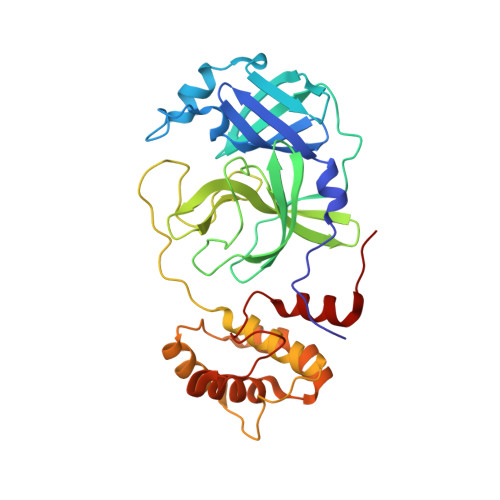
As an essential enzyme of SARS-CoV-2, the COVID-19 pathogen, main protease (M Pro ) is a viable target to develop antivirals for the treatment of COVID-19. By varying chemical compositions at both P2 and P3 positions and the N-terminal protection group, we synthesized 18 tripeptidyl M Pro inhibitors that contained also an aldehyde warhead and β-(S-2-oxopyrrolidin-3-yl)-alaninal at the P1 position. Systematic characterizations of these inhibitors were conducted, including their in vitro enzymatic inhibition potency, X-ray crystal structures of their complexes with M Pro , their inhibition of M Pro transiently expressed in 293T cells, and cellular toxicity and SARS-CoV-2 antiviral potency of selected inhibitors. These inhibitors have a large variation of determined in vitro enzymatic inhibition IC 50 values that range from 4.8 to 650 nM. The determined in vitro enzymatic inhibition IC 50 values reveal that relatively small side chains at both P2 and P3 positions are favorable for achieving high in vitro M Pro inhibition potency, the P3 position is tolerable toward unnatural amino acids with two alkyl substituents on the α-carbon, and the inhibition potency is sensitive toward the N-terminal protection group. X-ray crystal structures of M Pro bound with 16 inhibitors were determined. In all structures, the M Pro active site cysteine interacts covalently with the aldehyde warhead of the bound inhibitor to form a hemithioacetal that takes an S configuration. For all inhibitors, election density around the N-terminal protection group is weak indicating possible flexible binding of this group to M Pro . In M Pro , large structural variations were observed on residues N142 and Q189. Unlike their high in vitro enzymatic inhibition potency, most inhibitors showed low potency to inhibit M Pro that was transiently expressed in 293T cells. Inhibitors that showed high potency to inhibit M Pro transiently expressed in 293T cells all contain O-tert-butyl-threonine at the P3 position. These inhibitors also exhibited relatively low cytotoxicity and high antiviral potency. Overall, our current and previous studies indicate that O-tert-butyl-threonine at the P3 site is a key component to achieve high cellular and antiviral potency for tripeptidyl aldehyde inhibitors of M Pro .
- Texas A&M Drug Discovery Laboratory, Department of Chemistry, Texas A&M University, College Station, TX, 77843, USA.
Organizational Affiliation: