7RSO

AMC016 SOSIP.v4.2 in complex with PGV04 Fab


Experimental Data Snapshot

  • Method: ELECTRON MICROSCOPY
  • Resolution: 4.10 Å
  • Aggregation State: PARTICLE 
  • Reconstruction Method: SINGLE PARTICLE 

wwPDB Validation   3D Report Full Report


This is version 1.2 of the entry. See complete history


Literature

The Glycan Hole Area of HIV-1 Envelope Trimers Contributes Prominently to the Induction of Autologous Neutralization.

Schorcht, A.Cottrell, C.A.Pugach, P.Ringe, R.P.Han, A.X.Allen, J.D.van den Kerkhof, T.L.G.M.Seabright, G.E.Schermer, E.E.Ketas, T.J.Burger, J.A.van Schooten, J.LaBranche, C.C.Ozorowski, G.de Val, N.Bader, D.L.V.Schuitemaker, H.Russell, C.A.Montefiori, D.C.van Gils, M.J.Crispin, M.Klasse, P.J.Ward, A.B.Moore, J.P.Sanders, R.W.

(2022) J Virol 96: e0155221-e0155221

  • DOI: https://doi.org/10.1128/JVI.01552-21
  • Primary Citation of Related Structures:  
    7RSN, 7RSO

  • PubMed Abstract: 

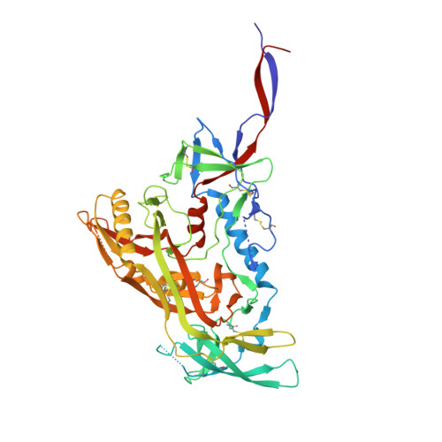
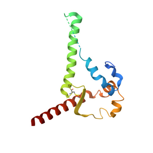
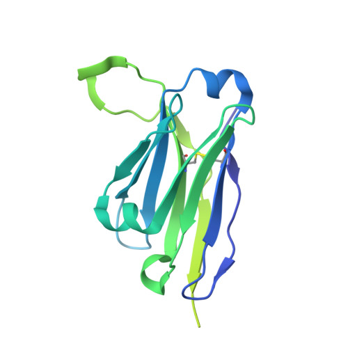
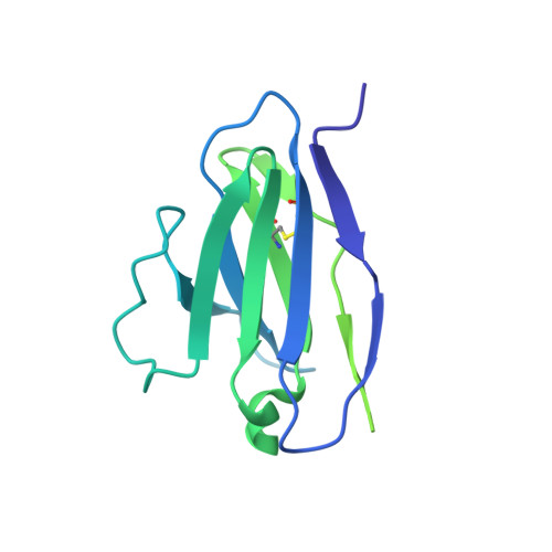
    The human immunodeficiency virus type 1 (HIV-1) trimeric envelope glycoprotein (Env) is heavily glycosylated, creating a dense glycan shield that protects the underlying peptidic surface from antibody recognition. The absence of conserved glycans, due to missing potential N-linked glycosylation sites (PNGS), can result in strain-specific, autologous neutralizing antibody (NAb) responses. Here, we sought to gain a deeper understanding of the autologous neutralization by introducing holes in the otherwise dense glycan shields of the AMC011 and AMC016 SOSIP trimers. Specifically, when we knocked out the N130 and N289 glycans, which are absent from the well-characterized B41 SOSIP trimer, we observed stronger autologous NAb responses. We also analyzed the highly variable NAb responses induced in rabbits by diverse SOSIP trimers from subtypes A, B, and C. Statistical analysis, using linear regression, revealed that the cumulative area exposed on a trimer by glycan holes correlates with the magnitude of the autologous NAb response. IMPORTANCE Forty years after the first description of HIV-1, the search for a protective vaccine is still ongoing. The sole target for antibodies that can neutralize the virus are the trimeric envelope glycoproteins (Envs) located on the viral surface. The glycoprotein surface is covered with glycans that shield off the underlying protein components from recognition by the immune system. However, the Env trimers of some viral strains have holes in the glycan shield. Immunized animals developed antibodies against such glycan holes. These antibodies are generally strain specific. Here, we sought to gain a deeper understanding of what drives these specific immune responses. First, we show that strain-specific neutralizing antibody responses can be increased by creating artificial holes in the glycan shield. Second, when studying a diverse set of Env trimers with different characteristics, we found that the surface area of the glycan holes contributes prominently to the induction of strain-specific neutralizing antibodies.


  • Organizational Affiliation

    Department of Medical Microbiology and Infection Prevention, Amsterdam Infection & Immunity Institute (AI&AII), Amsterdam UMC, Location Meibergdreef, University of Amsterdam, Amsterdam, The Netherlands.


Macromolecules
Find similar proteins by:  (by identity cutoff)  |  3D Structure
Entity ID: 1
MoleculeChains Sequence LengthOrganismDetailsImage
AMC016 gp120A,
E [auth C],
G [auth D]
486Human immunodeficiency virus 1Mutation(s): 0 
Entity Groups  
Sequence Clusters30% Identity50% Identity70% Identity90% Identity95% Identity100% Identity
Sequence Annotations
Expand
  • Reference Sequence
Find similar proteins by:  (by identity cutoff)  |  3D Structure
Entity ID: 2
MoleculeChains Sequence LengthOrganismDetailsImage
AMC016 gp41B,
F [auth E],
H [auth F]
154Human immunodeficiency virus 1Mutation(s): 0 
Entity Groups  
Sequence Clusters30% Identity50% Identity70% Identity90% Identity95% Identity100% Identity
Sequence Annotations
Expand
  • Reference Sequence
Find similar proteins by:  (by identity cutoff)  |  3D Structure
Entity ID: 3
MoleculeChains Sequence LengthOrganismDetailsImage
PGV04 Fab heavy chainC [auth H],
I [auth G],
K [auth I]
229Homo sapiensMutation(s): 0 
Entity Groups  
Sequence Clusters30% Identity50% Identity70% Identity90% Identity95% Identity100% Identity
Sequence Annotations
Expand
  • Reference Sequence
Find similar proteins by:  (by identity cutoff)  |  3D Structure
Entity ID: 4
MoleculeChains Sequence LengthOrganismDetailsImage
PGV04 kappa chainD [auth L],
J,
L [auth K]
208Homo sapiensMutation(s): 0 
Entity Groups  
Sequence Clusters30% Identity50% Identity70% Identity90% Identity95% Identity100% Identity
Sequence Annotations
Expand
  • Reference Sequence
Oligosaccharides

Help

Entity ID: 6
MoleculeChains Length2D Diagram Glycosylation3D Interactions
beta-D-mannopyranose-(1-4)-2-acetamido-2-deoxy-beta-D-glucopyranose-(1-4)-2-acetamido-2-deoxy-beta-D-glucopyranose
AA [auth a],
IA [auth i],
MA [auth m],
O,
UA [auth u],
AA [auth a],
IA [auth i],
MA [auth m],
O,
UA [auth u],
W
3N/AN-Glycosylation
Glycosylation Resources
GlyTouCan:  G15407YE
GlyCosmos:  G15407YE
GlyGen:  G15407YE
Entity ID: 7
MoleculeChains Length2D Diagram Glycosylation3D Interactions
alpha-D-mannopyranose-(1-2)-alpha-D-mannopyranose-(1-3)-[alpha-D-mannopyranose-(1-6)]beta-D-mannopyranose-(1-4)-2-acetamido-2-deoxy-beta-D-glucopyranose-(1-4)-2-acetamido-2-deoxy-beta-D-glucopyranoseBA [auth b],
NA [auth n],
P
6N/AN-Glycosylation
Glycosylation Resources
GlyTouCan:  G56014GC
GlyCosmos:  G56014GC
GlyGen:  G56014GC
Entity ID: 8
MoleculeChains Length2D Diagram Glycosylation3D Interactions
alpha-D-mannopyranose-(1-3)-[alpha-D-mannopyranose-(1-6)]beta-D-mannopyranose-(1-4)-2-acetamido-2-deoxy-beta-D-glucopyranose-(1-4)-2-acetamido-2-deoxy-beta-D-glucopyranoseCA [auth c],
OA [auth o],
Q
5N/AN-Glycosylation
Glycosylation Resources
GlyTouCan:  G22768VO
GlyCosmos:  G22768VO
GlyGen:  G22768VO
Small Molecules
Ligands 1 Unique
IDChains Name / Formula / InChI Key2D Diagram3D Interactions
NAG
Query on NAG

Download Ideal Coordinates CCD File 
AB [auth A]
AC [auth D]
BB [auth A]
BC [auth D]
CB [auth A]
AB [auth A],
AC [auth D],
BB [auth A],
BC [auth D],
CB [auth A],
CC [auth D],
DB [auth A],
DC [auth D],
EB [auth A],
EC [auth D],
FB [auth A],
FC [auth D],
GB [auth A],
GC [auth D],
HB [auth A],
HC [auth D],
IB [auth B],
IC [auth D],
JB [auth B],
JC [auth D],
KB [auth B],
KC [auth D],
LB [auth C],
LC [auth D],
MB [auth C],
MC [auth F],
NB [auth C],
NC [auth F],
OB [auth C],
OC [auth F],
PB [auth C],
QB [auth C],
RB [auth C],
SB [auth C],
TB [auth C],
UB [auth C],
VB [auth C],
WA [auth A],
WB [auth C],
XA [auth A],
XB [auth E],
YA [auth A],
YB [auth E],
ZA [auth A],
ZB [auth E]
2-acetamido-2-deoxy-beta-D-glucopyranose
C8 H15 N O6
OVRNDRQMDRJTHS-FMDGEEDCSA-N
Experimental Data & Validation

Experimental Data

  • Method: ELECTRON MICROSCOPY
  • Resolution: 4.10 Å
  • Aggregation State: PARTICLE 
  • Reconstruction Method: SINGLE PARTICLE 
EM Software:
TaskSoftware PackageVersion
RECONSTRUCTIONcryoSPARC2
MODEL REFINEMENTRosetta

Structure Validation

View Full Validation Report


Entry History & Funding Information

Deposition Data


Funding OrganizationLocationGrant Number
National Institutes of Health/National Institute Of Allergy and Infectious Diseases (NIH/NIAID)United StatesP01 AI110657
National Institutes of Health/National Institute Of Allergy and Infectious Diseases (NIH/NIAID)United StatesUM1 AI144462
National Institutes of Health/National Institute Of Allergy and Infectious Diseases (NIH/NIAID)United StatesF31 AI131873

Revision History  (Full details and data files)

  • Version 1.0: 2021-09-15
    Type: Initial release
  • Version 1.1: 2021-11-03
    Changes: Database references
  • Version 1.2: 2022-01-26
    Changes: Database references